Antitussive and Anti-inflammatory Dual-active Agents Developed from Natural Product Lead Compound 1-Methylhydantoin

 ,
, 
Abstract
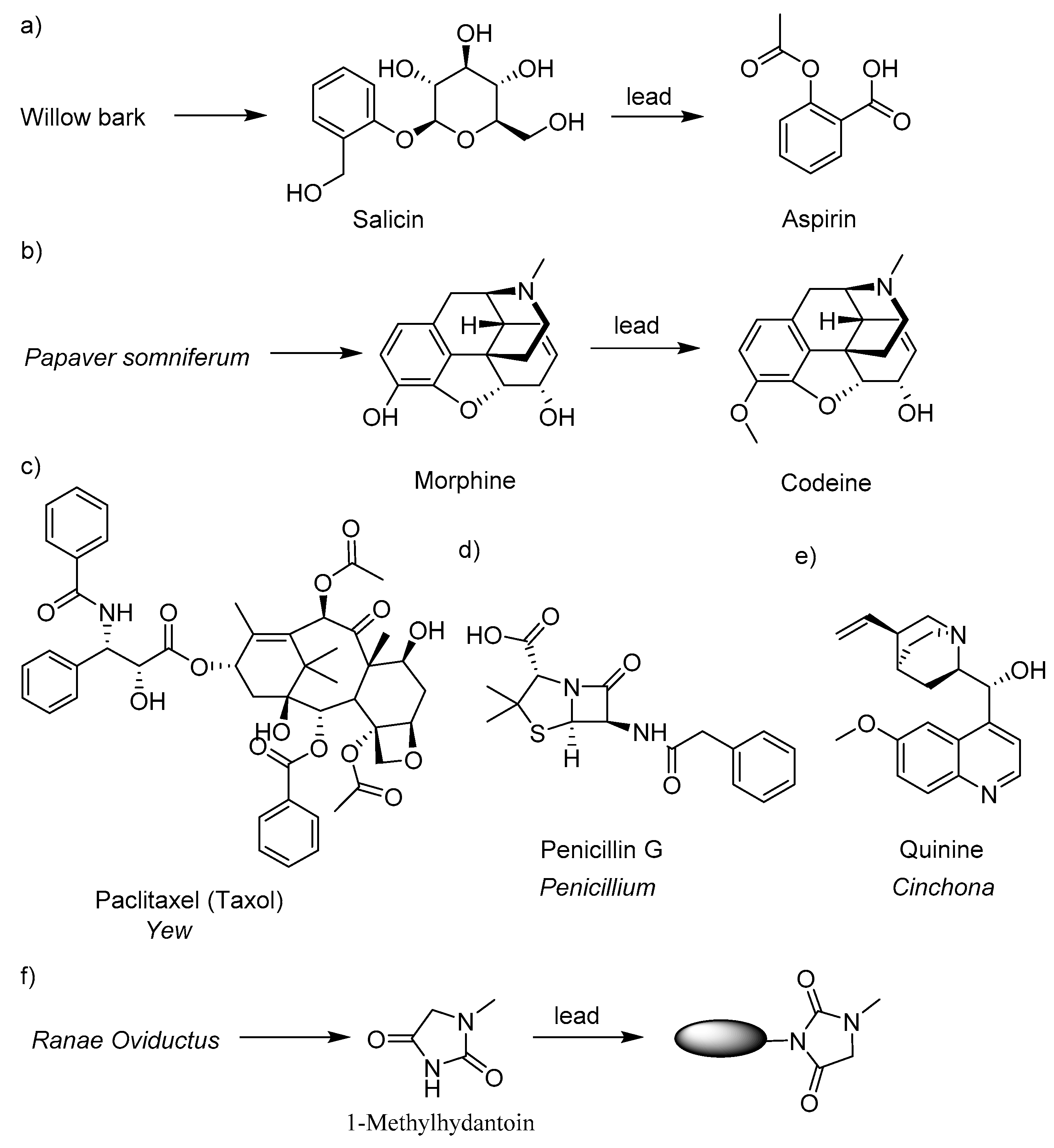
:1. Introduction
2. Result and Discussion
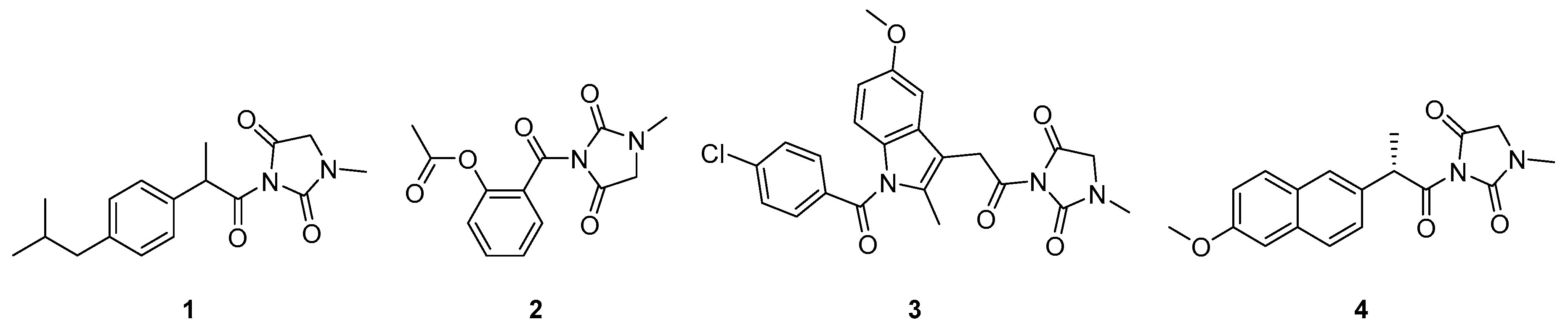
2.1. Molecular Design and Synthesis
2.2. Bioactivity Evaluation
3. Materials and Methods
3.1. Reagents and Instruments
3.2. Synthesis of 3-(2-(4-isobutylphenyl)propanoyl)-1-methylimidazolidine-2,4-dione (1)
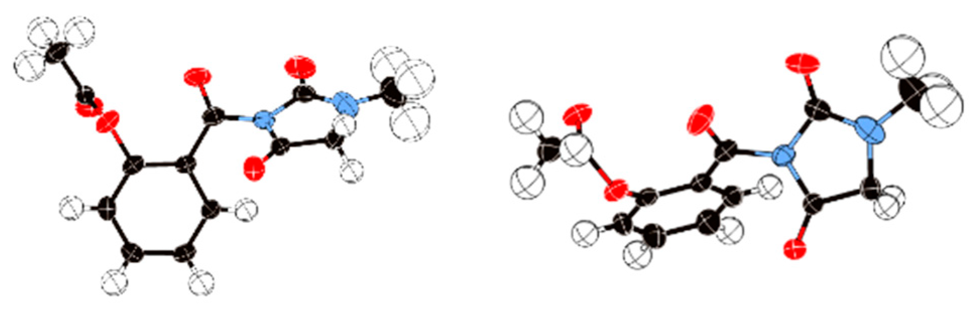
3.3. Synthesis of (2-(3-methyl-2,5-dioxoimidazolidine-1-carbonyl)phenyl acetate) (2)
3.4. Synthesis of 3-(2-(1-(4-chlorobenzoyl)-5-methoxy-2-methyl-1H-indol-3-yl)acetyl)-1-methylimidazolidine-2,4-dione (3)
3.5. Synthesis of ((S)-3-(2-(6-methoxynaphthalen-2-yl)propanoyl)-1-methylimidazolidine-2,4-dione) (4)
3.6. Animals
3.7. Antitussive Activity
3.8. Anti-Inflammatory Activity
3.9. Anti-PM 2.5 Acute Pneumonia Bioactivity
3.10. Statistical Analysis
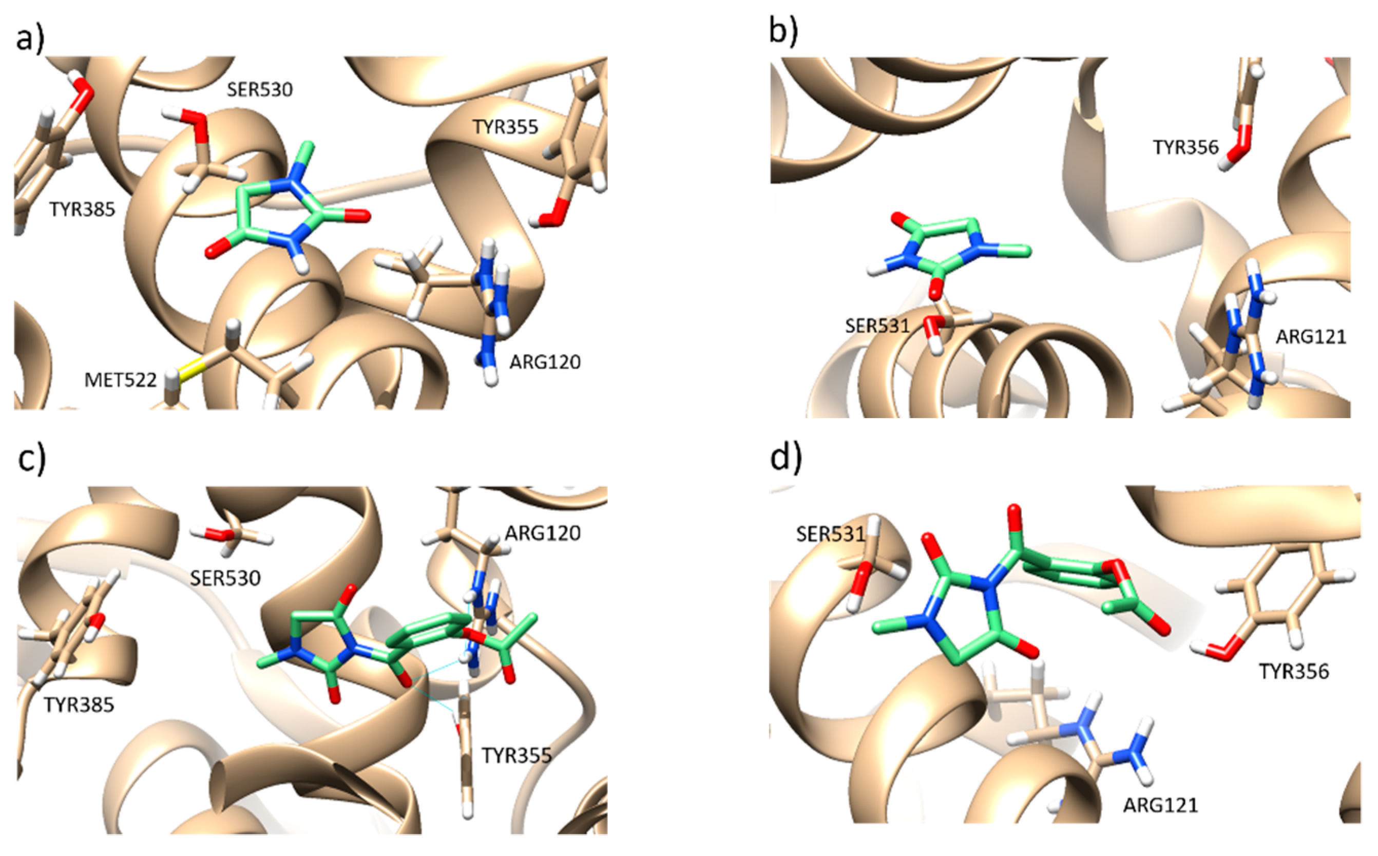
3.11. In Silico Study
4. Conclusions
Supplementary Materials
Author Contributions
Funding
Conflicts of Interest
References
- Patwardhan, B.; Vaidya, A.D.B.; Chorghade, M. Ayurveda and natural products drug discovery. Curr. Sci. 2004, 86, 789–799. [Google Scholar]
- Gu, J.; Gui, Y.; Chen, L.; Yuan, G.; Lu, H.-Z.; Xu, X. Use of natural products as chemical library for drug discovery and network pharmacology. PLoS ONE 2013, 8, e62839. [Google Scholar] [CrossRef] [PubMed]
- Dias, D.A.; Urban, S.; Roessner, U. A historical overview of natural products in drug discovery. Metabolites 2012, 2, 303–336. [Google Scholar] [CrossRef] [PubMed]
- Ho, T.T.; Tran, Q.T.N.; Chai, C.L.L. The polypharmacology of natural products. Future Med. Chem. 2018, 10, 1361–1368. [Google Scholar] [CrossRef] [PubMed]
- Olğaç, A.; Orhan, I.E.; Banoglu, E. The potential role of in silico approaches to identify novel bioactive molecules from natural resources. Future Med. Chem. 2017, 9, 1665–1686. [Google Scholar] [CrossRef]
- Sagandykova, G.N.; Pomastowski, P.P.; Kaliszan, R.; Buszewski, B. Modern analytical methods for consideration of natural biological activity. Trac-Trends Anal. Chem. 2018, 109, 198–213. [Google Scholar] [CrossRef]
- Sun, H.H.; Liu, Z.H.; Zhao, H.M.; Ang, E.L. Recent advances in combinatorial biosynthesis for drug discovery. Drug Des. Dev. Ther. 2015, 9, 823–833. [Google Scholar]
- Yang, G.X.; Ma, G.L.; Li, H.; Huang, T.; Xiong, J.; Hu, J.F. Advanced natural products chemistry research in china between 2015 and 2017. Chin. J. Nat. Med. 2018, 16, 881–906. [Google Scholar] [CrossRef]
- Newman, D.J.; Cragg, G.M. Natural products as sources of new drugs over the last 25 years. J. Nat. Prod. 2007, 70, 461–477. [Google Scholar] [CrossRef]
- Lascano, S.; Lopez, M.; Arimondo, P.B. Natural products and chemical biology tools: Alternatives to target epigenetic mechanisms in cancers. Chem. Rec. 2018, 18, 1854–1876. [Google Scholar] [CrossRef]
- Noleto-Dias, C.; Ward, J.L.; Bellisai, A.; Lomax, C.; Beale, M.H. Salicin-7-sulfate: A new salicinoid from willow and implications for herbal medicine. Fitoterapia 2018, 127, 166–172. [Google Scholar] [CrossRef] [PubMed]
- Li, Q.L.; Zhang, H.B. Research progress on the synthesis of morphine alkaloids. Chin. J. Org. Chem. 2017, 37, 1629–1652. [Google Scholar] [CrossRef]
- Khanna, C.; Rosenberg, M.; Vail, D.M. A review of paclitaxel and novel formulations including those suitable for use in dogs. J. Vet. Intern. Med. 2015, 29, 1006–1012. [Google Scholar] [CrossRef] [PubMed]
- Kim, H.J.; Joo, H.G. Paclitaxel inhibits the hyper-activation of spleen cells by lipopolysaccharide and induces cell death. J. Vet. Sci. 2016, 17, 453–458. [Google Scholar] [CrossRef] [PubMed] [Green Version]
- Kim, J.H.; Lee, J.O.; Kim, N.; Lee, H.J.; Lee, Y.W.; Kim, H.I.; Kim, S.J.; Park, S.H.; Kim, H.S. Paclitaxel suppresses the viability of breast tumor mcf7 cells through the regulation of ef1 alpha and foxo3a by ampk signaling. Int. J. Oncol. 2015, 47, 1874–1880. [Google Scholar] [CrossRef] [PubMed]
- Barreiro, C.; Martin, J.F.; Garcia-Estrada, C. Proteomics shows new faces for the old penicillin producer penicillium chrysogenum. J. Biomed. Biotechnol. 2012, 2012, 105109. [Google Scholar] [CrossRef]
- Atanasov, A.G.; Waltenberger, B.; Pferschy-Wenzig, E.M.; Linder, T.; Wawrosch, C.; Uhrin, P.; Temml, V.; Wang, L.M.; Schwaiger, S.; Heiss, E.H.; et al. Discovery and resupply of pharmacologically active plant-derived natural products: A review. Biotechnol. Adv. 2015, 33, 1582–1614. [Google Scholar] [CrossRef] [PubMed] [Green Version]
- Harvey, A.L. Natural products in drug discovery. Drug Discov. Today 2008, 13, 894–901. [Google Scholar] [CrossRef]
- Shen, B. A new golden age of natural products drug discovery. Cell 2015, 163, 1297–1300. [Google Scholar] [CrossRef]
- Wang, S.; Xu, Y.; Wang, Y.; Yang, H.; Lv, Z.; Jin, X.; Wang, Y. Simultaneous determination of six active components in Oviductus Ranae via quantitative analysis of multicomponents by single marker. J. Anal. Methods Chem. 2017, 2017, 9194847. [Google Scholar] [CrossRef]
- Wang, S.; Xu, Y.; Wang, Y.; Lv, Z.; Cui, Q.; Jin, X.; Wang, Y. HPLC fingerprint combined with quantitation of main effective components and chemometrics as an efficient method for quality evaluation of Oviductus Ranae. Nat. Prod. Commun. 2017, 12, 1495–1499. [Google Scholar] [CrossRef]
- Xu, Y.; Wang, S.-h.; Luo, Y.; Yang, H.-l.; Hu, X.; Wang, Y.-s.; Qu, X.-b. Separation of steroidal constituents of Oviductus Ranae by one-step method high-speed counter-current chromatography. J. Liq. Chromatogr. Relat. Technol. 2015, 38, 1494–1498. [Google Scholar] [CrossRef]
- Xu, Y.; Wang, S.-H.; Luo, Y.; Wang, Y.-S.; Qu, X.-B. Evaluation of the merits of the new method of Oviductus Ranae by HPLC-DAD. J. Liq. Chromatogr. Relat. Technol. 2015, 38, 1218–1222. [Google Scholar] [CrossRef]
- Gan, Y.; Xiao, Y.; Wang, S.; Guo, H.; Liu, M.; Wang, Z.; Wang, Y. Protein-based fingerprint analysis for the identification of Oviductus Ranae using RP-HPLC. Molecules 2019, 24, 1687. [Google Scholar] [CrossRef] [PubMed]
- Zhang, Y.; Zhu, K.; Cui, H.; Liu, Y.; Lu, Y.-F.; Pan, H.-W.; Zhao, H.-P.; Qi, L.; Yang, X.-D.; Zhou, H.-L. Toxicological evaluation of Oviductus Ranae: Acute, sub-acute and genotoxicity studies in mice and rats. J. Ethnopharmacol. 2017, 203, 101–109. [Google Scholar] [CrossRef] [PubMed]
- Nogueira, B.A.; Ildiz, G.O.; Henriques, M.S.C.; Paixao, J.A.; Fausto, R. Structural and spectroscopic characterization of the second polymorph of 1-methylhydantoin. J. Mol. Struct. 2017, 1148, 111–118. [Google Scholar] [CrossRef]
- Lu, H.B.; Kong, D.J.; Wu, B.; Wang, S.H.; Wang, Y.S. Synthesis and evaluation of anti-inflammatory and antitussive activity of hydantion derivatives. Lett. Drug Des. Discovery 2012, 9, 638–642. [Google Scholar] [CrossRef]
- Bauer, A.; Bronstrupt, M. Industrial natural product chemistry for drug discovery and development. Nat. Prod. Rep. 2014, 31, 35–60. [Google Scholar] [CrossRef]
- Chaudhari, P.S.; Chitlange, S.S.; Nanda, R.K. Synthesis and biological evaluation of novel 2-(4-acetyl-3-methyl- 5-(arylamino) thiophen-2-yl)-3-arylquinazolin-4(3h)-one derivatives as potential anti-inflammatory and antioxidant agents. Anti-Inflamm. Anti-Allergy Agents Med. Chem. 2018, 17, 102–114. [Google Scholar] [CrossRef]
- Ouellet, M.; Riendeau, D.; Percival, M.D. A high level of cyclooxygenase-2 inhibitor selectivity is associated with a reduced interference of platelet cyclooxygenase-1 inactivation by aspirin. Proc. Natl. Acad. Sci. USA 2001, 98, 14583–14588. [Google Scholar] [CrossRef] [Green Version]
- Cristina, A.; Leonte, D.; Vlase, L.; Bencze, L.C.; Imre, S.; Marc, G.; Apan, B.; Mogoșan, C.; Zaharia, V. Heterocycles 48. Synthesis, characterization and biological evaluation of imidazo[2,1-b][1,3,4]thiadiazole derivatives as anti-inflammatory agents. Molecules 2018, 23, 2425. [Google Scholar] [CrossRef] [PubMed]
- Zidar, N.; Odar, K.; Glavac, D.; Jerse, M.; Zupanc, T.; Stajer, D. Cyclooxygenase in normal human tissues--is cox-1 really a constitutive isoform, and cox-2 an inducible isoform? J. Cell. Mol. Med. 2009, 13, 3753–3763. [Google Scholar] [CrossRef] [PubMed]
- Jackson, L.M.; Wu, K.C.; Mahida, Y.R.; Jenkins, D.; Hawkey, C.J. Cyclooxygenase (cox) 1 and 2 in normal, inflamed, and ulcerated human gastric mucosa. Gut 2000, 47, 762–770. [Google Scholar] [CrossRef] [PubMed]
- Gomes, A.; Fernandes, E.; Silva, A.M.S.; Pinto, D.; Santos, C.M.M.; Cavaleiro, J.A.S.; Lima, J. Anti-inflammatory potential of 2-styrylchromones regarding their interference with arachidonic acid metabolic pathways. Biochem. Pharmacol. 2009, 78, 171–177. [Google Scholar] [CrossRef] [PubMed]
- Sharma, V.; Bhatia, P.; Alam, O.; Javed Naim, M.; Nawaz, F.; Ahmad Sheikh, A.; Jha, M. Recent advancement in the discovery and development of cox-2 inhibitors: Insight into biological activities and sar studies (2008–2019). Bioorg. Chem. 2019, 89, 103007. [Google Scholar] [CrossRef] [PubMed]
- Anderson, G.D.; Hauser, S.D.; McGarity, K.L.; Bremer, M.E.; Isakson, P.C.; Gregory, S.A. Selective inhibition of cyclooxygenase (cox)-2 reverses inflammation and expression of cox-2 and interleukin 6 in rat adjuvant arthritis. J. Clin. Investig. 1996, 97, 2672–2679. [Google Scholar] [CrossRef] [PubMed]
- Ouyang, Y.F.; Yang, H.; Zhang, P.; Wang, Y.; Kaur, S.; Zhu, X.L.; Wang, Z.; Sun, Y.T.; Hong, W.; Ngeow, Y.F.; et al. Synthesis of 2,4-diaminopyrimidine core-based derivatives and biological evaluation of their anti-tubercular activities. Molecules 2017, 22, 1592. [Google Scholar] [CrossRef]
- Lin, X.; Chai, L.; Liu, B.M.; Chen, H.L.; Zheng, L.; Liu, Q.; Lin, C.W. Synthesis, biological evaluation, and docking studies of a novel sulfonamido-based gallate as pro-chondrogenic agent for the treatment of cartilage. Molecules 2017, 22, 3. [Google Scholar] [CrossRef]
- Lucido, M.J.; Orlando, B.J.; Vecchio, A.J.; Malkowski, M.G. Crystal structure of aspirin-acetylated human cyclooxygenase-2: Insight into the formation of products with reversed stereochemistry. Biochemistry 2016, 55, 1226–1238. [Google Scholar] [CrossRef]
- Corazzi, T.; Leone, M.; Maucci, R.; Corazzi, L.; Gresele, P. Direct and irreversible inhibition of cyclooxygenase-1 by nitroaspirin (ncx 4016). J. Pharmacol. Exp. Ther. 2005, 315, 1331–1337. [Google Scholar] [CrossRef]
- Wang, D.; Wang, S.; Chen, X.; Xu, X.; Zhu, J.; Nie, L.; Long, X. Antitussive, expectorant and anti-inflammatory activities of four alkaloids isolated from bulbus of fritillaria wabuensis. J. Ethnopharmacol. 2012, 139, 189–193. [Google Scholar] [CrossRef] [PubMed]
- Tubaro, A.; Dri, P.; Delbello, G.; Zilli, C.; Loggia, R.D. The croton oil ear test revisited. Agents Actions 1986, 17, 347–349. [Google Scholar] [CrossRef] [PubMed]
- Yue, J.; Zhang, H.; Cai, Z.; Zhao, Y.; Ye, W.; Xuan, Z.; Ying, L.; Qin, Y.; Gu, M.; Jin, J. Bufei huoxue capsule attenuates pm2.5-induced pulmonary inflammation in mice. Evid.-Based Complement Altern. Med. 2017, 2017, 1575793. [Google Scholar]
- Sidhu, R.S.; Lee, J.Y.; Yuan, C.; Smith, W.L. Comparison of cyclooxygenase-1 crystal structures: Cross-talk between monomers comprising cyclooxygenase-1 homodimers. Biochemistry 2010, 49, 7069–7079. [Google Scholar] [CrossRef] [PubMed]
- Orlando, B.J.; Lucido, M.J.; Malkowski, M.G. The structure of ibuprofen bound to cyclooxygenase-2. J. Struct. Biol. 2015, 189, 62–66. [Google Scholar] [CrossRef] [PubMed]
Sample Availability: Not available. |


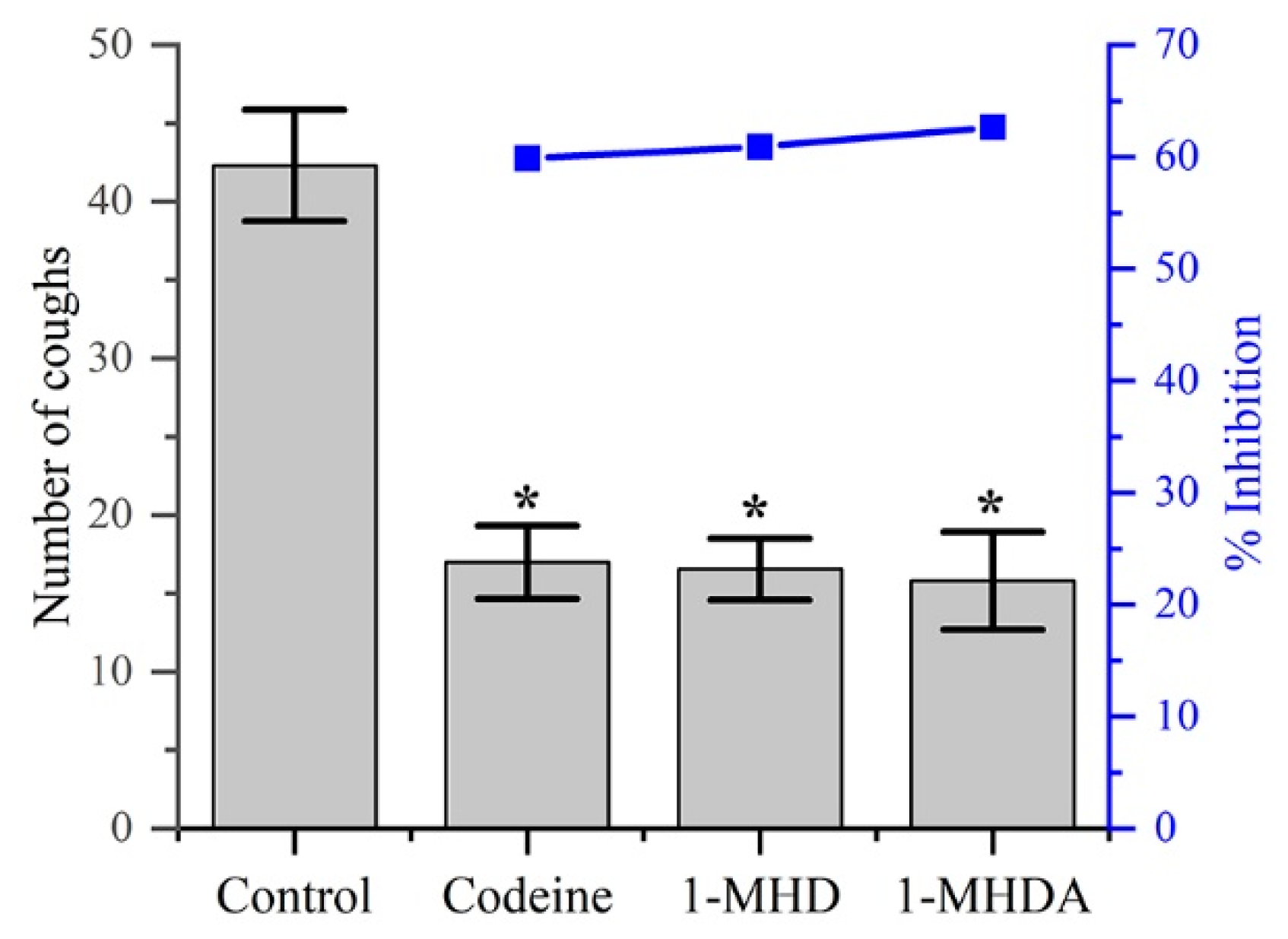
| Group | Dose (mg/Kg) | No. of Animals | Latent Period Cough (s) | No. of Coughs | Inhibition (%) |
|---|---|---|---|---|---|
| Control | 5% CMC | 10 | 27.20 ± 5.73 | 42.30 ± 3.55 | - |
| Codeine phosphate | 30 | 10 | 51.50 ± 5.41* | 16.98 ± 2.33* | 59.86 |
| 1-MHD | 100 | 10 | 38.20 ± 9.69* | 16.54 ± 1.96* | 60.90 |
| 2 | 100 | 10 | 41.00 ± 4.25* | 15.80 ± 3.12* | 62.65 |
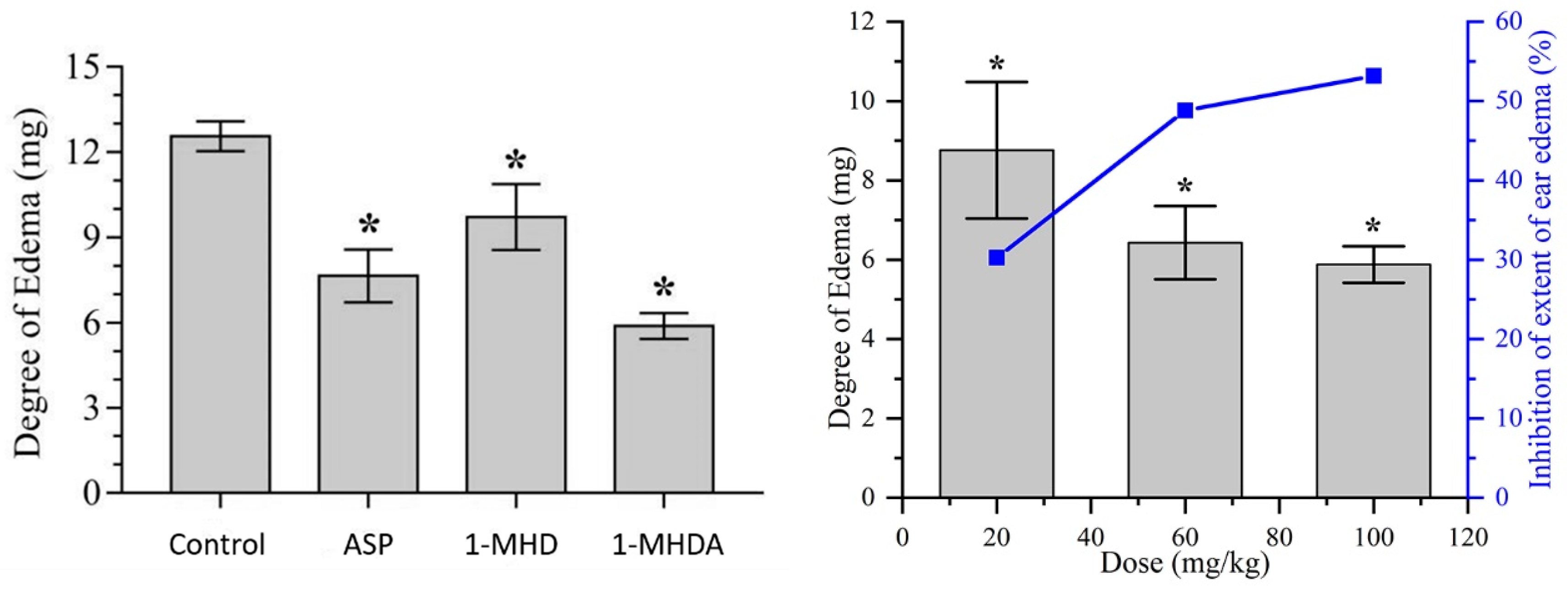
| Group | Dose (mg/Kg) | No. of Animals | Degree of Edema (mg) | Inhibition (%) |
|---|---|---|---|---|
| Control | 5% CMC | 10 | 12.56 ± 0.52 | - |
| ASP | 100 | 10 | 7.65 ± 0.93* | 39.09 |
| 1-MHD | 100 | 10 | 9.71 ± 1.16* | 22.69 |
| 2 | 100 (high dose) | 10 | 5.88 ± 0.46* | 53.18 |
| 2 | 60 (medium dose) | 10 | 6.43 ± 0.92* | 48.81 |
| 2 | 20 (low dose) | 10 | 8.76 ± 1.72* | 30.25 |
© 2019 by the authors. Licensee MDPI, Basel, Switzerland. This article is an open access article distributed under the terms and conditions of the Creative Commons Attribution (CC BY) license (http://creativecommons.org/licenses/by/4.0/).
Share and Cite
Xu, Y.; Wang, F.; Guo, H.; Wang, S.; Ni, S.; Zhou, Y.; Wang, Z.; Bao, H.; Wang, Y. Antitussive and Anti-inflammatory Dual-active Agents Developed from Natural Product Lead Compound 1-Methylhydantoin. Molecules 2019, 24, 2355. https://doi.org/10.3390/molecules24132355
Xu Y, Wang F, Guo H, Wang S, Ni S, Zhou Y, Wang Z, Bao H, Wang Y. Antitussive and Anti-inflammatory Dual-active Agents Developed from Natural Product Lead Compound 1-Methylhydantoin. Molecules. 2019; 24(13):2355. https://doi.org/10.3390/molecules24132355
Chicago/Turabian StyleXu, Yang, Fang Wang, Hongye Guo, Shihan Wang, Shuling Ni, Yan Zhou, Zhihan Wang, Huiwei Bao, and Yongsheng Wang. 2019. "Antitussive and Anti-inflammatory Dual-active Agents Developed from Natural Product Lead Compound 1-Methylhydantoin" Molecules 24, no. 13: 2355. https://doi.org/10.3390/molecules24132355