
Anti-Apoptotic Effects of Carotenoids in Neurodegeneration


Department of Human Nutrition and Hospitality Management, College of Human Environmental Sciences, The University of Alabama, Tuscaloosa, AL 35487, USA
*
Author to whom correspondence should be addressed.
Molecules 2020, 25(15), 3453; https://doi.org/10.3390/molecules25153453
Submission received: 6 July 2020
/
Revised: 27 July 2020
/
Accepted: 27 July 2020
/
Published: 29 July 2020
(This article belongs to the Special Issue Neuroprotective Potential of Bioactive Natural Compounds in Oxidative Stress Conditions)
Abstract
:Apoptosis, programmed cell death type I, is a critical part of neurodegeneration in cerebral ischemia, Parkinson’s, and Alzheimer’s disease. Apoptosis begins with activation of pro-death proteins Bax and Bak, release of cytochrome c and activation of caspases, loss of membrane integrity of intracellular organelles, and ultimately cell death. Approaches that block apoptotic pathways may prevent or delay neurodegenerative processes. Carotenoids are a group of pigments found in fruits, vegetables, and seaweeds that possess antioxidant properties. Over the last several decades, an increasing number of studies have demonstrated a protective role of carotenoids in neurodegenerative disease. In this review, we describe functions of commonly consumed carotenoids including lycopene, β-carotene, lutein, astaxanthin, and fucoxanthin and their roles in neurodegenerative disease models. We also discuss the underlying cellular mechanisms of carotenoid-mediated neuroprotection, including their antioxidant properties, role as signaling molecules, and as gene regulators that alleviate apoptosis-associated brain cell death.
1. Introduction
Apoptosis, programmed cell death type I, naturally occurs during development and maturation of the healthy brain [1,2]. However, apoptotic pathways also play an important role in brain-associated diseases that are caused by loss of functional populations of neurons. During neurotoxic stimulation, such as oxidative stress, excitotoxicity, or neuroinflammation, brain cells undergo death receptor-mediated (extrinsic) apoptosis, mitochondria-mediated (intrinsic) apoptosis, or a combination of both. Activated death receptors such as tumor necrosis factor receptor 1 (TNFR1) form a trimer, recruiting adapter proteins such as TNFR1-associated death domain protein (TRADD), TNRF-associated factor (TRAF) 2, cellular inhibitor of apoptosis (cIAP) 1 and 2, and Fas-associated death domain protein (FADD). FADD further recruits procaspase 8 and converts it to its active form. Death receptor-mediated caspase 8 activation results in cleavage of Bcl-2 interacting protein (Bid), a pro-apoptotic BH3-only protein, producing truncated Bid (tBid) [3]. tBid is translocated to the mitochondrial membrane and recruits multidomain proapoptotic Bcl-2 proteins such as Bcl-2-associated X protein (Bax). Bax is oligomerized with itself or other pro-apoptotic proteins such as Bak, making the mitochondrial membrane permeable (Figure 1).
Unlike BH3-only and multidomain pro-apoptotic proteins, B-cell lymphoma-2 (Bcl-2), and B-cell lymphoma-extra large (Bcl-xL) contain a BH4 domain, a key domain with anti-apoptotic activities. There, anti-apoptotic Bcl-2 family proteins bind directly to pro-apoptotic Bcl-2 proteins preventing oligomerization. However, excess activation of pro-apoptotic Bcl-2 proteins causes loss of mitochondrial membrane integrity leading to the release of cytochrome c. In healthy mitochondria, cytochrome c is found on the inner membrane and acts as an electron carrier in the electron transport chain. However, during mitochondrial outer membrane permeabilization, cytochrome c is released into the cytoplasm due to its hydrophilicity. Cytoplasmic cytochrome c interacts with apoptotic protease activating factor (Apaf) 1 and dATP to form an apoptosome. The apoptosome activates caspase 9, which further activates executor caspases such as caspase 3 that degrade functional and structural proteins in the cell. This ultimately leads to neuronal death. Although opening of the mitochondrial permeability transition pore (mtPTP), a large mitochondrial inner membrane death channel [4,5,6], occurs in both apoptotic and necrotic neurons, it may be regulated by Bcl-2 family proteins. Bcl-xL binds directly to the c-subunit of the F1Fo ATP synthase [7], a voltage-sensitive channel that acts like a mtPTP [8,9], preventing proton leak through the pore. Bcl-xL is reported to form a complex with other mtPTP candidates such as the voltage-dependent anion channel and adenine nucleotide translocator to prevent mitochondrial dysfunction [10,11].
Enhanced neuronal apoptosis after cerebral ischemia is well documented in both rodent models of stroke (focal cerebral ischemia) and cardiac arrest (global cerebral ischemia) [12,13,14,15,16,17]. In vitro models that mimic cerebral ischemia including treatment with glutamate, hydrogen peroxide, hypoxia, and oxygen-glucose deprivation consistently show increased levels of apoptosis [18,19,20,21]. Although there are numerous studies demonstrating involvement of apoptotic pathways during ischemic insult, key findings indicate that cerebral ischemia (1) alters the proportion of pro-apoptotic vs. anti-apoptotic Bcl-2 proteins, (2) increases mitochondrial membrane permeability, and (3) activates caspases in both mitochondria-dependent and independent manners.
Apoptosis also plays an important role in Alzheimer’s disease [22,23,24]. Accumulation of amyloid-β induces both death receptor-mediated extrinsic and mitochondria-mediated intrinsic apoptosis. For example, TNFR1 is necessary during amyloid-β-induced neuronal death [25], and amyloid-β increases both mRNA and protein levels of Fas ligand [26]. In addition, treatment with amyloid-β manipulates expression of Bcl-2 family proteins and induces cytochrome c release from mitochondria [27,28,29,30]. Amyloid-β-mediated activation of initiator caspases including caspase 8 [31,32] and executor caspases like caspase 3 [33] has been widely investigated. Caspases carry out proteolytic cleavage of amyloid precursor protein (APP) producing amyloid-β [34,35,36]. Aggregation of tau, the neurofibrillary tangles found in both Alzheimer’s and Parkinson’s-affected brains, is regulated by caspases. The Asp421 residue on the C-terminal of tau is subjected to caspases, and increased proteolytic cleavage of tau is found in apoptotic neurons [37,38]. Therefore, apoptosis is involved in both initiation and propagation of the pathology of Alzheimer’s disease.
Loss of dopaminergic neurons in the substantia nigra results in Parkinson’s disease. Although autophagy and necrosis are also involved during neurodegeneration, apoptotic features including increased abundance of pro-apoptotic Bcl-2 protein, activation of caspases, and cytochrome c release are found during progression of Parkinson’s disease [39,40,41]. Mitochondrial dysfunction, particularly impairment of complex I, is a key feature of Parkinson’s pathology [42,43,44]. Currently, application of complex I inhibitors like 1-methyl-4-phenyl-1,2,3,6-tetrahydropyridine (MPTP) and rotenone are widely used to mimic Parkinsonian pathology in research [45,46]. Additionally, mutations of PARK genes or abnormality of PARK gene products is highly associated with mitochondria-mediated neuronal damage. Oligomerization of α-synuclein, encoded by the PARK1 gene, triggers apoptosis [47]. Depletion of Parkin or PINK1, encoded by the PARK2 and PARK6 genes, respectively, impairs removal of dysfunctional mitochondria and increases cellular reactive oxygen species (ROS) load [48]. DJ-1, encoded by the PARK7 gene, binds directly to the F1Fo ATP synthase and anti-apoptotic Bcl-xL to improve mitochondrial energy metabolism and dopaminergic neuron survival [49].
Application of pharmacological inhibitors and genetic modifications that block oligomerization of pro-apoptotic Bcl-2 proteins [50,51] or enhance the function of Bcl-2 or Bcl-xL protect neurons from injury [12,18,52,53]. Treatment with caspase inhibitors has protective effects in cerebral ischemia and neurodegeneration models [18,54,55,56]. However, there are significant barriers to applying molecular-based strategies to treat brain-associated diseases in humans. In recent decades, an increasing number of researchers have highlighted the potential significance of nutrients and dietary phytochemicals as neuroprotectants [57,58,59,60]. Since neurodegeneration occurs gradually over a number of years, improving life-style factors like diet may delay apoptotic neuronal loss.
2. Overview of Carotenoids
Carotenoids are a family of yellow, orange, and red pigments found in plants, fungi, bacteria, and algae. Carotenoids play an important role in photosynthesis by extending the range of wavelengths of light that drive the reaction forward. In addition, carotenoids protect photosynthetic organisms from damage caused by excessive exposure to light [61,62]. There are over 600 types of carotenoids found in nature that provide a large range of health benefits. Carotenoids can be categorized into six different groups according to their chemical structures: hydrocarbons, hydroxycarotenoids, epoxycarotenoids, aldehydes, ketones, and carboxylic acids [63]. The many types of carotenoids are categorized into two large groups, the carotenes and the xanthophylls based on their chemical components. Carotenes consist of lycopene, β-carotene, and α-carotene based on their structure containing only a parent hydrocarbon chain [64]. Xanthophylls consist of structures that contain oxygen as a functional group such as beta-cryptoxanthin, neoxanthin, astaxanthin, canthaxanthin, zeaxanthin, fucoxanthin, and lutein [64].
A large number of carotenoids are present in the human diet, with foods such as fruits and vegetables containing rich pigmentation. It has also been noted that nearly 20 types of carotenoids are present in the blood and tissues of the human body [65]. Experimentation with supplementation of carotenoids can affect cellular redox status, gene expression, immune responses, cell growth, and development [66]. Studies are continuing to be conducted to fully understand the extensive effects carotenoids have on the brain and brain-related diseases.
2.1. Lycopene
Lycopene is a carotenoid responsible for the red-pink color of certain vegetables and fruits, including tomatoes, grapefruit, watermelon, and guava. Chemically, it is a hydrocarbon carotenoid, composed strictly of carbon and hydrogen assembled from eight isoprene units. In the fresh fruit and vegetable form, lycopene exists predominantly as trans-lycopene. Upon exposure to heat, trans-lycopene undergoes isomerization to cis-lycopene [67]. Numerous clinical studies have demonstrated the greater absorption and bioavailability of cis-versus trans-lycopene [68,69,70]. Bioavailability remains stable throughout heat treatment [71,72], allowing lycopene to retain its biological function during food processing. Extensive research has been published on lycopene, and it has potential benefits in regard to cancer, cardiovascular disease, and inflammatory disease [73]. The efficiency of lycopene crossing the blood–brain barrier remains controversial. Oral administration of lycopene (10, 30, and 50 mg/kg) increases lycopene levels in liver, the adrenals, spleen, lymph nodes, and intestinal tissues in dogs, but lycopene was not detected in the brain [74]. However, oral administration of lycopene (8 mg/kg) increased the lycopene concentration in mouse brain, and improvement of this delivery method by using a lycopene-loaded microemulsion significantly enhanced brain distribution of lycopene [75]. Lycopene acts as an antioxidant with greater singlet oxygen quenching abilities than other carotenoids such as β-carotene, lutein, and zeaxanthin [76]. Application of lycopene has been shown to protect brain cells from oxidative stress-induced damage. Treatment with lycopene inhibits both lipid peroxidation of membranes and accumulation of hydrogen peroxide and superoxide in both in vitro and in vivo neurodegeneration models [77,78,79,80,81]. Interestingly, pro-oxidant effects of lycopene have been reported under co-treatment with a lipid-soluble radical generator in fibroblasts [82], thus, the efficacy of lycopene as an antioxidant may differ by both oxidant and cell type. Additionally, lycopene upregulates intracellular antioxidant defense system components including superoxide dismutase, catalase, glutathione peroxidase, and glutathione [79,81,83]. The antioxidant properties of lycopene are particularly important in protecting mitochondria [77,80,83]. Mitochondria are the central oxygen consuming organelle by virtue of housing the electron transport chain, but mitochondria are also responsible for production of ROS [84]. Excess ROS accumulation and impaired antioxidant defenses result in mitochondrial dysfunction, and mitochondrial dysfunction results in depletion of energy, initiation of apoptosis, and ultimately neuronal death in the brain [85,86]. Treatment with lycopene prevents loss of mitochondrial inner membrane potential during ROS challenge [78,83,87]. Qu et al. show that lycopene improves energy metabolism in primary cortical neurons by preventing loss of complex I, II, III, and IV activity during amyloid-β treatment [77]. This same group also suggests that lycopene is capable of protecting mitochondrial DNA that encodes NADH dehydrogenase subunit 6 and cytochrome c oxidase subunit I, key subunits found in complexes I and IV, respectively [77]. Although not yet confirmed, lycopene-mediated energy retention may occur via protection of other mitochondrial DNA like the ATP6 and ATP8 genes, which encode the a-subunit of F1Fo ATP synthase (Complex V), the enzyme complex responsible for ATP production [88].
Anti-apoptotic functions of lycopene in neurodegeneration have been reported relatively recently. Lycopene prevents loss of anti-apoptotic proteins including Bcl-2 and Bcl-xL [80,89], while inhibiting pro-apoptotic proteins like Bax [78,80] under neurotoxic conditions. Lycopene protects primary hippocampal neurons from apoptotic death by blocking opening of mtPTP [90]. Neurons treated with lycopene have decreased mitochondrial depolarization and cytochrome c release, indicating retention of mitochondrial membrane integrity [77,83]. Pathological concentrations of intracellular calcium trigger opening of mtPTP [91]. Zhang et al. showed that treatment with lycopene increases activity of the Ca2+-ATPase and Ca2+-Mg2+-ATPase, transport proteins that assist with clearance of calcium from the cell [79]. Thus, lycopene may prevent mtPTP-associated neuronal death by regulating calcium homeostasis. In addition, lycopene prevents activation of caspase 3 [78]. Although further investigation is required to elucidate the exact molecular mechanism behind lycopene-mediated inhibition of apoptosis, lycopene may also regulate the transcription of genes that control apoptosis [78,79].
Treatment with lycopene increases phosphorylation of Akt [78,79]. Akt, also called protein kinase B, is phosphorylated upon activation of phosphoinositide 3-kinase (PI3K), and phosphorylated Akt regulates transcription factors (Figure 1) such as nuclear factor erythroid 2-related factor 2 (Nrf2) and NF-κB, influencing gene expression. Indeed, lycopene is shown to manipulate Nrf2 and NF-κB targets, alleviating oxidative stress or inflammation-associated damage in brain cells [80,92]. Lycopene mediates the functionality of the cyclooxygenase-2 (COX-2) pathway, which is primarily responsible for production of pro-inflammatory prostaglandins [93,94], and lycopene reduces the release of inflammatory cytokines and chemokines including interleukin (IL)-6 and IL-8 and acute phase proteins such as C-reactive protein (CRP) [95,96]. Lycopene has been shown to attenuate cognitive deficits by reducing inflammation in the gut–liver–brain axis as well as improving glycolipid metabolism [97]. Recent studies suggest that anti-inflammatory effects of lycopene may help alleviate neuropsychiatric diseases such as post-traumatic stress disorder and depression [98,99]. Treatment with lycopene improves stress-associated behavior in rodents by preventing accumulation of inflammatory cytokines in the brain [98,99]. Additionally, Akt was previously shown to upregulate expression of the Bcl-2 gene by activating the cAMP-response element (CRE) [100], and Nrf2 [101] bind directly to the Bcl-x gene, contributing to the retention of protective Bcl-2 and Bcl-xL. Lycopene also activates the mitogen-activated protein kinase (MAPK)/extracellular signal-regulated kinase (Erk) pathway in the brain [102]. The MAPK/Erk pathway (Figure 1) includes a series of kinase activities including a serine/threonine-specific protein kinase Raf, mitogen-activated protein kinase kinase (MEK/MAPKK), and Erk. Phosphorylated Erk translocates into the nucleus and activates transcription factors such as Elk-1 and Msk regulating synaptic plasticity [103,104].
Lycopene treatment in vitro and in vivo using intracerebroventricular administration show improved production of neurotrophic factors including brain-derived neurotrophic factor (BDNF) and nerve growth factor (NGF) during neurotoxic challenge [78,105]. BDNF blocks neuronal apoptosis during excitotoxicity by inducing phosphorylation of Akt at Ser473 and Thr308 sites [106,107]. Akt is shown to upregulate genes containing CRE such as Bcl-2 [100,106]. NGF also plays a role in regulating apoptosis via activation of PI3K/Akt and MAPK/Erk pathways [2,108]. Interaction between NGF and tropomyosin receptor kinase A (TrkA) activates Erk signaling to phosphorylate the BH3-only protein Bim, and this inactivates the pro-apoptotic function of Bim [109,110].
2.2. β-Carotene
β-carotene is a carotenoid exhibiting the red and orange pigments in certain fruits and vegetables such as carrots, sweet potatoes, and squash. It contains an unsaturated hydrocarbon chain with β-rings at both ends. Symmetric cleavage of β-carotene by β-carotene-15,15-oxygenase releases two molecules of retinal, an aldehyde form of vitamin A [111,112,113]. Retinal is further metabolized to retinoic acid or to retinol and retinyl ester, an acid, alcohol, and ester form of vitamin A, respectively [111,112]. Therefore, β-carotene is an important provitamin A. Nuclear receptors including retinoic acid receptor (RAR) and retinoid X receptor (RXR) are dimerized and regulate genes containing retinoid acid response elements. Studies suggest that RAR and RXR may be required during neurogenesis and for neuroprotection against apoptosis [114,115].
β-carotene decreases accumulation of ROS including hydrogen peroxide and lipid peroxide radicals [116,117]. β-carotene is characterized as a direct acting low molecular weight antioxidant due to its scavenger nature. The ability of β-carotene to work against lipid peroxidation and decrease oxidative stress makes it an important factor in brain-related diseases. [118]. Lower levels of β-carotene in the cerebral cortex are associated with traumatic brain injury [119]. Unlike vitamin A, which can produce oxidative stress with excess treatment, β-carotene does not show pro-oxidant activity in the brain, and it has been thus suggested as a safer antioxidant [116]. Treatment with β-carotene improves retention of intracellular antioxidants such as glutathione and superoxide dismutase during neurotoxicity [118,120]. β-carotene increases protein levels of nuclear Nrf2 and upregulates downstream targets containing antioxidant response elements (ARE) such as NAD(P)H quinone dehydrogenase and heme oxygenase-1 [120]. Oxidative stress is a major contributor to neuronal apoptosis during neurotoxicity, thus, treatment with β-carotene may prevent neuronal loss in ROS-associated brain diseases. Indeed, β-carotene is shown to interfere with apoptotic pathways in the brain. Treatment with β-carotene prevents loss of Bcl-2 and accumulation of Bax and caspase 3 in a mouse model of traumatic brain injury [120].
In addition to its protection against oxidative stress, β-carotene may play a beneficial role during neuronal development. β-carotene improves cognitive and social behavior of newborn mice [121]. Oral supplementation of β-carotene increases levels of BDNF in mice [121,122]. Interaction between N-terminal cleaved mature BDNF (129–247 amino acid residues) and a receptor like tropomyosin receptor kinase B (TrkB) activates PI3K/Akt pathways, and this interaction activates cAMP response element-binding protein (CREB), a transcription factor that regulates genes that control neuronal development and plasticity [123,124]. N-terminal (19–128 amino acids) containing proBDNF binds to the P75 neurotrophin receptor, a type of tumor necrosis factor (TNF) receptor that mediates apoptosis, indicating that β-carotene-mediated neuronal development and survival may be via regulation of mature BDNF/proBDNF in the brain.
2.3. Astaxanthin
Astaxanthin is a red-orange carotenoid found naturally in algae, yeast, salmon, trout, krill, shrimp, and crayfish [125]. It is a xanthophyll carotenoid made up of carbon, hydrogen, and oxygen [126]. Chemically, it consists of two terminal rings joined by a polyene chain, with a hydroxyl group and a carbonyl group located on each terminal ring [126]. The polyene chain consists of conjugated double bonds that result in delocalized electrons that can be readily donated and thus provide astaxanthin with strong antioxidant capabilities [127]. Studies have shown astaxanthin as possessing anti-lipid peroxidation [128], anti-inflammatory [129], anti-diabetic [130], and anti-cancer activity [131], as well as cardiovascular disease prevention [132] and immuno-modulation [133]. Astaxanthin does have a low oral bioavailability, but absorption is enhanced by combined consumption with fats [134]. Its lipophilic nature allows it to cross the blood–brain barrier, and all-trans-astaxanthin is detected in rat hippocampus and cortex after oral administration of astaxanthin (100 mg/kg) [135]. Astaxanthin has been shown to exert anti-oxidative protective effects in neurological disorders and is shown to decrease markers of lipid oxidation including malodialdehyde, decrease protein levels of ROS-generating enzymes such as NADPH oxidase 4 and p22phox protein, and increase antioxidant enzymes such as superoxide dismutase [136]. Astaxanthin was also shown to increase protein levels of Nrf2 and p62, thereby protecting against oxidative stress [136,137]. Nrf2 is normally sequestered in the cytosol bound to Keap1 [138]. Oxidative stress and an increase in p62, which competes to bind to Keap1 [139], cause Nrf2 to translocate to the nucleus to activate ARE [138]. Astaxanthin also protects against apoptosis by regulating mitochondrial proteins [136,137,140,141]. Treatment with astaxanthin increased levels of Bcl-2 [136,140,141] and decreased levels of Bax and cleaved caspase 3 [136,137,140,141]. By increasing levels of Bcl-2, astaxanthin prevents the release of cytochrome c into the cytosol and thereby prevents apoptosis [142]. Activated Akt has been shown to phosphorylate Bad, and this prevents the activation of caspase 9 and caspase 3 to protect mitochondria [143]. Treatment with astaxanthin prevents loss of mitochondrial membrane potential in rat hippocampal neurons [140].
Astaxanthin also appears to exert protective effects via anti-inflammatory pathways [136,137,141]. The NF-κB pathway is a pro-inflammatory pathway that regulates the production of cytokines and chemokines [144]. While in an inactive state, NF-κB binds to inhibitor of nuclear factor kappa B (IκBα) and is sequestrated in the cytoplasm. However, while in an active state, IκBα is dissociated from NF-κB, and NF-κB is translocated to the nucleus, regulating gene expression. Treatment with astaxanthin decreases protein levels of total and nuclear NF-κB in the hippocampus and cortex [136,141]. TNF-α [145], IL-1β [146], and IL-6 [147] are all pro-inflammatory cytokines produced by macrophages and are involved in the NF-κB pathway, and TNF-α and IL-1β have both been shown to activate NF-κB [144] and thus can operate in a positive feedback loop. In models of brain injury and disease, treatment with astaxanthin attenuated the increase in levels of TNF-α, IL-1β, and IL-6, exerting an anti-inflammatory effect [136,137,141]. Administration of astaxanthin also attenuates depressive-like behaviors in rodents by preventing neuroinflammation [148,149,150] and regulating survival pathways including PI3K/Akt [151,152]. Decrease in cytokine levels may be due to astaxanthin inhibiting invasion of macrophages, the source of cytokine production [136]. Persistent elevation of inflammatory cytokines has been shown to lead to demyelination and eventually cell death [153], and thus by lowering levels of pro-inflammatory cytokines astaxanthin protects against neuronal cell death. In addition, Zang et al. also suggest that astaxanthin prevents swelling of brain cells by downregulating the Na+-K+-2Cl− co-transporter-1 (NKCC1), a key transporter implicated in brain edema, via the NF-κB pathway [141]. Inhibition of NKCC1 has been shown to attenuate disruption of the blood–brain barrier and thus provide a neuroprotective effect [154].
2.4. Lutein
Lutein is a plant-originated carotenoid found in green-leafy vegetables such as spinach, kale, broccoli, and other foods including eggs and avocados. Lutein contains an unsaturated hydrocarbon chain with cyclic structures at each end and is isomeric with zeaxanthin. Along with zeaxanthin, it is the only carotenoid that is found in the macula of the eye after crossing the blood–retina barrier [155]. Treatment with lutein attenuates retinopathy associated with photo stress, inflammation, and oxidative stress [156,157,158], and dietary lutein prevents aging- and diabetes-related eye diseases [159,160]. Lutein also crosses the blood–brain barrier to allow for delivery to various regions of the brain. Lutein is detected in the cortex, cerebellum, striatum, and hippocampus of rhesus monkeys after oral administration at 0.25–0.5 μmol/kg [161,162]. Therefore, studies investigating lutein’s benefits in brain development and potential protective effects during neurodegeneration are quickly becoming relevant [163,164].
Lutein decreases accumulation of lipid hydroperoxides and formation of malondialdehyde and thiobarbituric acid adduct, markers of lipid peroxidation, and it also inhibits oxygen photo-consumption in a concentration-dependent manner [165]. Lutein restores other antioxidants including glutathione, superoxide dismutase, and catalase in the brain during ischemic challenge [166]. Interestingly, Mohn et al. show that lutein levels in the mitochondrial membrane are inversely associated with fatty acid oxidation in the primate brain [162], indicating a potential protective role of lutein in brain pathology associated with mitochondrial dysfunction. Treatment with lutein significantly decreases mitochondrial ROS in Caenorhabditis elegans (Caenorhabditis elegans) lacking nuo-5/NDUFS1 and lpd-5/NDUFS4, genes that encode subunits of complex I in the electron transport chain [167]. This study demonstrates that depletion of complex I decreases mitochondrial ATP production, increases mitochondria ROS accumulation, and increases lipid peroxidation leading to synapse dysfunction, whereas treatment with lutein reverses these effects to rescue cholinergic synapse activity [167]. Lutein increases Nrf2 and decreases NF-κB subunit p65, thus, lutein may regulate transcription of genes that control antioxidant activity and inflammation [168,169,170,171,172].
Treatment with lutein protects neurons and brain-derived cells from apoptotic death. Lutein prevents loss of Bcl-2 and Bcl-xL, accumulation of Bax, and activation of caspases 3 and 8 in brain disease models of cerebral ischemia and Parkinson’s disease [172,173,174]. Caspase 3 is a central protease responsible for destruction of cellular structure and execution of apoptosis. In addition, caspase 3 disables functions of pro-survival proteins. Caspase 3 carries out post-translational cleavage of Bcl-xL and Bcl-2 into pro-death molecules [18,19,175,176,177]. Therefore, lutein-mediated caspase 3 inactivation may support the maintenance of full-length Bcl-xL and Bcl-2 and their anti-apoptotic properties in neurons. Furthermore, lutein may effectively reverse apoptosis triggered during glutamate-mediated excitotoxicity. During cerebral ischemia and neurodegeneration, excess glutamate activates N-methyl-d-aspartate (NMDA) receptors allowing a surge of intracellular calcium. This surge activates destructive enzymes including calpain and phospholipases to cause neuronal death. Lutein prevents loss of retinal ganglion cells against NMDA challenge [174] and suppresses death signaling including MAPK-mediated c-Jun activation [172,174]. In addition to apoptosis, Fung et al. showed that hypoxia-induced AMP activated protein kinase (AMPK) inhibits mTOR, followed by induction of microtubule-associated protein 1A/1B-light chain 3 (LC3) II, treatment with lutein protects glial cells by reversing this pathway [173]. LC3-I is the cytosolic form and LC3-II is the phosphatidylethanolamine conjugated form found in autophagosomes. Elevated ROS are shown to enhance recruitment of LC3 [178], therefore, both downregulation of LC3-II and attenuation of ROS in lutein treatment may prevent uncontrolled autophagic degradation of brain cells.
2.5. Fucoxanthin
Fucoxanthin is a carotenoid found in brown seaweed [179,180] with antioxidant and anti-inflammatory properties. Although functions of fucoxanthin are less researched compared to other carotenoids, an increasing number of studies suggest potential anti-apoptotic roles of fucoxanthin in neurodegenerative diseases [181,182,183,184]. Pharmacokinetic analysis of fucoxanthin and its metabolite fucoxanthinol in plasma after intragastric administration of fucoxanthin in rats has been performed [185,186], and tissue distribution of fucoxanthin after oral administration has also been performed using mouse models [187]. However, it is unknown if fucoxanthin crosses the blood–brain barrier. Despite limited information on fucoxanthin and the blood-brain barrier permeability, experiments have been performed that show that animals who undergo middle cerebral artery occlusion (MCAO) demonstrate brain tissue damage associated with apoptosis and neurological deficits, but fucoxanthin treatment reverses this MCAO-induced brain injury [188]. Treatment with fucoxanthin improves cognitive and motor function in animals challenged by neurodegeneration and ischemic insults [183,188,189]. Intragastric and intracerebroventricular administration of fucoxanthin reverses apoptosis in mouse traumatic brain injury and ischemic stroke models via enhancing the abundance of Bcl-2 and decreasing Bax, cytochrome c release, and caspase 3 activity [184,188]. Neuroblastoma SH-SY5Y cells treated with fucoxanthin show decreased numbers of pyknotic nuclei and condensation of chromatin, indicators of apoptosis, during ROS and amyloid-β challenge [181,182].
Fucoxanthin and its metabolites have strong radical scavenging activity [190]. For example, hydroxyl radical scavenging activity of fucoxanthin is 13.5 times greater than that of α-tocopherol [190]. In addition to scavenging ROS, fucoxanthin may directly occupy the docking site, altering protein function [191]. Fucoxanthin potentially binds directly to amyloid-β, preventing formation of pathological amyloid-β fibril [191]. Fucoxanthin also regulates intracellular redox status by regulating antioxidant-sensitive genes. Studies show that fucoxanthin activates transcription factor Nrf2 [184,188] and redox sensitive targets of Nrf2 such as heme oxygenase-1, superoxide dismutase, and Bcl-2 expression, protecting rodent brain against neurotoxic insult [184,188]. Fucoxanthin fails to reverse apoptosis in mice lacking Nrf2, indicating that Nrf2 is a critical part of fucoxanthin-mediated neuroprotection [184,188]. Fucoxanthin exhibits protective properties via activation of the PI3K/Akt pathway during neurotoxic stimulation [181,182]. PI3K/Akt promotes nuclear translocation of Nrf2 [192], thus, the PI3K/Akt pathway is a likely upstream component of fucoxanthin-mediated upregulation of antioxidant genes.
3. Conclusions
Neurons in the mature brain are generally postmitotic, and thus, maintaining viable neuron populations is critically important to support brain function and activity. During neurodegeneration, activation of apoptotic signaling causes structural and functional deterioration of the brain. To date, there is no treatment to cure neurodegenerative disease pathology. As an alternative to post-damage treatment, it is important to find ways to prevent neuronal apoptosis and alleviate apoptosis-induced cellular damage. In this review, we describe inhibitory roles of various carotenoids against neuronal apoptosis (Table 1). Carotenoids act as antioxidants to block upstream triggers of apoptosis and ROS-associated mitochondrial dysfunction. Carotenoids thus prevent energy deficit and subsequent activation of mitochondria-derived apoptotic molecules. In addition, carotenoids influence numerous transcription factors such as Nrf2, and NF-κB to regulate genes for apoptosis, antioxidant defense, and inflammation. Although we highlight roles of carotenoids in apoptotic signaling in this review, carotenoids may also intervene in other death pathways such as necroptosis, pyroptosis, and ferroptosis. Therefore, future investigation to distinguish the specific molecular mechanisms of neuronal death that are involved and continued translational studies in human subjects will be invaluable in outlining protective and therapeutic roles of carotenoids in neurodegeneration.
Author Contributions
Writing and editing: H.-A.P., M.M.H., S.B., J.J., and K.M.C.-W. All authors have read and agreed to the published version of the manuscript.
Funding
This work was supported by RG14811 (University of Alabama) and A-52248–217101–100 (University of Alabama).
Conflicts of Interest
The authors declare no conflict of interest.
References
- Yamaguchi, Y.; Miura, M. Programmed cell death in neurodevelopment. Dev. Cell 2015, 32, 478–490. [Google Scholar] [CrossRef] [PubMed] [Green Version]
- Kristiansen, M.; Ham, J. Programmed cell death during neuronal development: The sympathetic neuron model. Cell Death Differ. 2014, 21, 1025–1035. [Google Scholar] [CrossRef] [PubMed] [Green Version]
- Galluzzi, L.; Blomgren, K.; Kroemer, G. Mitochondrial membrane permeabilization in neuronal injury. Nat. Rev. Neurosci. 2009, 10, 481–494. [Google Scholar] [CrossRef] [PubMed]
- Martin, L.J.; Semenkow, S.; Hanaford, A.; Wong, M. Mitochondrial permeability transition pore regulates Parkinson’s disease development in mutant alpha-synuclein transgenic mice. Neurobiol. Aging 2014, 35, 1132–1152. [Google Scholar] [CrossRef] [Green Version]
- Ludtmann, M.H.R.; Angelova, P.R.; Horrocks, M.H.; Choi, M.L.; Rodrigues, M.; Baev, A.Y.; Berezhnov, A.V.; Yao, Z.; Little, D.; Banushi, B.; et al. alpha-synuclein oligomers interact with ATP synthase and open the permeability transition pore in Parkinson’s disease. Nat. Commun. 2018, 9, 2293. [Google Scholar] [CrossRef] [Green Version]
- Mnatsakanyan, N.; Jonas, E.A. ATP synthase c-subunit ring as the channel of mitochondrial permeability transition: Regulator of metabolism in development and degeneration. J. Mol. Cell. Cardiol. 2020, 144, 109–118. [Google Scholar] [CrossRef]
- Alavian, K.N.; Li, H.; Collis, L.; Bonanni, L.; Zeng, L.; Sacchetti, S.; Lazrove, E.; Nabili, P.; Flaherty, B.; Graham, M.; et al. Bcl-xL regulates metabolic efficiency of neurons through interaction with the mitochondrial F1FO ATP synthase. Nat. Cell Biol. 2011, 13, 1224–1233. [Google Scholar] [CrossRef]
- Alavian, K.N.; Beutner, G.; Lazrove, E.; Sacchetti, S.; Park, H.A.; Licznerski, P.; Li, H.; Nabili, P.; Hockensmith, K.; Graham, M.; et al. An uncoupling channel within the c-subunit ring of the F1FO ATP synthase is the mitochondrial permeability transition pore. Proc. Natl. Acad. Sci. USA 2014, 111, 10580–10585. [Google Scholar] [CrossRef] [Green Version]
- Mnatsakanyan, N.; Llaguno, M.C.; Yang, Y.; Yan, Y.; Weber, J.; Sigworth, F.J.; Jonas, E.A. A mitochondrial megachannel resides in monomeric F1FO ATP synthase. Nat. Commun. 2019, 10, 5823. [Google Scholar] [CrossRef]
- Monaco, G.; Decrock, E.; Arbel, N.; van Vliet, A.R.; La Rovere, R.M.; De Smedt, H.; Parys, J.B.; Agostinis, P.; Leybaert, L.; Shoshan-Barmatz, V.; et al. The BH4 domain of anti-apoptotic Bcl-XL, but not that of the related Bcl-2, limits the voltage-dependent anion channel 1 (VDAC1)-mediated transfer of pro-apoptotic Ca2+ signals to mitochondria. J. Biol. Chem. 2015, 290, 9150–9161. [Google Scholar] [CrossRef] [Green Version]
- Gross, A.; Katz, S.G. Non-apoptotic functions of BCL-2 family proteins. Cell Death Differ. 2017, 24, 1348–1358. [Google Scholar] [CrossRef] [PubMed]
- Ofengeim, D.; Chen, Y.B.; Miyawaki, T.; Li, H.; Sacchetti, S.; Flannery, R.J.; Alavian, K.N.; Pontarelli, F.; Roelofs, B.A.; Hickman, J.A.; et al. N-terminally cleaved Bcl-xL mediates ischemia-induced neuronal death. Nat. Neurosci. 2012, 15, 574–580. [Google Scholar] [CrossRef] [Green Version]
- Park, H.A.; Kubicki, N.; Gnyawali, S.; Chan, Y.C.; Roy, S.; Khanna, S.; Sen, C.K. Natural vitamin E alpha-tocotrienol protects against ischemic stroke by induction of multidrug resistance-associated protein 1. Stroke 2011, 42, 2308–2314. [Google Scholar] [CrossRef] [PubMed] [Green Version]
- Park, H.A.; Licznerski, P.; Alavian, K.N.; Shanabrough, M.; Jonas, E.A. Bcl-xL Is Necessary for Neurite Outgrowth in Hippocampal Neurons. Antioxid. Redox Signal. 2015, 22, 93–108. [Google Scholar] [CrossRef] [PubMed] [Green Version]
- Miyawaki, T.; Mashiko, T.; Ofengeim, D.; Flannery, R.J.; Noh, K.M.; Fujisawa, S.; Bonanni, L.; Bennett, M.V.; Zukin, R.S.; Jonas, E.A. Ischemic preconditioning blocks BAD translocation, Bcl-xL cleavage, and large channel activity in mitochondria of postischemic hippocampal neurons. Proc. Natl. Acad. Sci. USA 2008, 105, 4892–4897. [Google Scholar] [CrossRef] [Green Version]
- Ferrer, I.; Planas, A.M. Signaling of cell death and cell survival following focal cerebral ischemia: Life and death struggle in the penumbra. J. Neuropathol. Exp. Neurol. 2003, 62, 329–339. [Google Scholar] [CrossRef] [Green Version]
- Broughton, B.R.; Reutens, D.C.; Sobey, C.G. Apoptotic mechanisms after cerebral ischemia. Stroke 2009, 40, e331–e339. [Google Scholar] [CrossRef] [Green Version]
- Park, H.A.; Licznerski, P.; Mnatsakanyan, N.; Niu, Y.; Sacchetti, S.; Wu, J.; Polster, B.M.; Alavian, K.N.; Jonas, E.A. Inhibition of Bcl-xL prevents pro-death actions of DeltaN-Bcl-xL at the mitochondrial inner membrane during glutamate excitotoxicity. Cell Death Differ. 2017, 24, 1963–1974. [Google Scholar] [CrossRef] [Green Version]
- Park, H.A.; Mnatsakanyan, N.; Broman, K.; Davis, A.U.; May, J.; Licznerski, P.; Crowe-White, K.M.; Lackey, K.H.; Jonas, E.A. Alpha-Tocotrienol Prevents Oxidative Stress-Mediated Post-Translational Cleavage of Bcl-xL in Primary Hippocampal Neurons. Int. J. Mol. Sci. 2019, 21, 220. [Google Scholar] [CrossRef] [Green Version]
- Malagelada, C.; Xifro, X.; Minano, A.; Sabria, J.; Rodriguez-Alvarez, J. Contribution of caspase-mediated apoptosis to the cell death caused by oxygen-glucose deprivation in cortical cell cultures. Neurobiol. Dis. 2005, 20, 27–37. [Google Scholar] [CrossRef]
- Wang, X.; Li, J.; Wu, D.; Bu, X.; Qiao, Y. Hypoxia promotes apoptosis of neuronal cells through hypoxia-inducible factor-1alpha-microRNA-204-B-cell lymphoma-2 pathway. Exp. Biol. Med. 2016, 241, 177–183. [Google Scholar] [CrossRef] [PubMed] [Green Version]
- Calissano, P.; Matrone, C.; Amadoro, G. Apoptosis and in vitro Alzheimer disease neuronal models. Commun. Integr. Biol. 2009, 2, 163–169. [Google Scholar] [CrossRef] [PubMed]
- Tatton, W.G.; Chalmers-Redman, R.; Brown, D.; Tatton, N. Apoptosis in Parkinson’s disease: Signals for neuronal degradation. Ann. Neurol. 2003, 53 (Suppl. 3), S61–S70. [Google Scholar] [CrossRef]
- Behl, C. Apoptosis and Alzheimer’s disease. J. Neural Transm. 2000, 107, 1325–1344. [Google Scholar] [CrossRef] [PubMed]
- He, P.; Zhong, Z.; Lindholm, K.; Berning, L.; Lee, W.; Lemere, C.; Staufenbiel, M.; Li, R.; Shen, Y. Deletion of tumor necrosis factor death receptor inhibits amyloid beta generation and prevents learning and memory deficits in Alzheimer’s mice. J. Cell Biol. 2007, 178, 829–841. [Google Scholar] [CrossRef] [PubMed] [Green Version]
- Morishima, Y.; Gotoh, Y.; Zieg, J.; Barrett, T.; Takano, H.; Flavell, R.; Davis, R.J.; Shirasaki, Y.; Greenberg, M.E. Beta-amyloid induces neuronal apoptosis via a mechanism that involves the c-Jun N-terminal kinase pathway and the induction of Fas ligand. J. Neurosci. 2001, 21, 7551–7560. [Google Scholar] [CrossRef] [PubMed]
- Kim, H.S.; Lee, J.H.; Lee, J.P.; Kim, E.M.; Chang, K.A.; Park, C.H.; Jeong, S.J.; Wittendorp, M.C.; Seo, J.H.; Choi, S.H.; et al. Amyloid beta peptide induces cytochrome C release from isolated mitochondria. Neuroreport 2002, 13, 1989–1993. [Google Scholar] [CrossRef]
- Yao, M.; Nguyen, T.V.; Pike, C.J. Beta-amyloid-induced neuronal apoptosis involves c-Jun N-terminal kinase-dependent downregulation of Bcl-w. J. Neurosci. 2005, 25, 1149–1158. [Google Scholar] [CrossRef]
- Selznick, L.A.; Zheng, T.S.; Flavell, R.A.; Rakic, P.; Roth, K.A. Amyloid beta-induced neuronal death is bax-dependent but caspase-independent. J. Neuropathol. Exp. Neurol. 2000, 59, 271–279. [Google Scholar] [CrossRef] [Green Version]
- Paradis, E.; Douillard, H.; Koutroumanis, M.; Goodyer, C.; LeBlanc, A. Amyloid beta peptide of Alzheimer’s disease downregulates Bcl-2 and upregulates bax expression in human neurons. J. Neurosci. 1996, 16, 7533–7539. [Google Scholar] [CrossRef] [Green Version]
- Ivins, K.J.; Thornton, P.L.; Rohn, T.T.; Cotman, C.W. Neuronal apoptosis induced by beta-amyloid is mediated by caspase-8. Neurobiol. Dis. 1999, 6, 440–449. [Google Scholar] [CrossRef] [PubMed] [Green Version]
- Rehker, J.; Rodhe, J.; Nesbitt, R.R.; Boyle, E.A.; Martin, B.K.; Lord, J.; Karaca, I.; Naj, A.; Jessen, F.; Helisalmi, S.; et al. Caspase-8, association with Alzheimer’s Disease and functional analysis of rare variants. PLoS ONE 2017, 12, e0185777. [Google Scholar] [CrossRef] [Green Version]
- Marin, N.; Romero, B.; Bosch-Morell, F.; Llansola, M.; Felipo, V.; Roma, J.; Romero, F.J. Beta-amyloid-induced activation of caspase-3 in primary cultures of rat neurons. Mech. Ageing Dev. 2000, 119, 63–67. [Google Scholar] [CrossRef]
- Lu, D.C.; Rabizadeh, S.; Chandra, S.; Shayya, R.F.; Ellerby, L.M.; Ye, X.; Salvesen, G.S.; Koo, E.H.; Bredesen, D.E. A second cytotoxic proteolytic peptide derived from amyloid beta-protein precursor. Nat. Med. 2000, 6, 397–404. [Google Scholar] [CrossRef] [PubMed]
- Stone, J.R.; Okonkwo, D.O.; Dialo, A.O.; Rubin, D.G.; Mutlu, L.K.; Povlishock, J.T.; Helm, G.A. Impaired axonal transport and altered axolemmal permeability occur in distinct populations of damaged axons following traumatic brain injury. Exp. Neurol. 2004, 190, 59–69. [Google Scholar] [CrossRef]
- Gervais, F.G.; Xu, D.; Robertson, G.S.; Vaillancourt, J.P.; Zhu, Y.; Huang, J.; LeBlanc, A.; Smith, D.; Rigby, M.; Shearman, M.S.; et al. Involvement of caspases in proteolytic cleavage of Alzheimer’s amyloid-beta precursor protein and amyloidogenic A beta peptide formation. Cell 1999, 97, 395–406. [Google Scholar] [CrossRef] [Green Version]
- Canu, N.; Dus, L.; Barbato, C.; Ciotti, M.T.; Brancolini, C.; Rinaldi, A.M.; Novak, M.; Cattaneo, A.; Bradbury, A.; Calissano, P. Tau cleavage and dephosphorylation in cerebellar granule neurons undergoing apoptosis. J. Neurosci. 1998, 18, 7061–7074. [Google Scholar] [CrossRef]
- Gamblin, T.C.; Chen, F.; Zambrano, A.; Abraha, A.; Lagalwar, S.; Guillozet, A.L.; Lu, M.; Fu, Y.; Garcia-Sierra, F.; LaPointe, N.; et al. Caspase cleavage of tau: Linking amyloid and neurofibrillary tangles in Alzheimer’s disease. Proc. Natl. Acad. Sci. USA 2003, 100, 10032–10037. [Google Scholar] [CrossRef] [Green Version]
- Vila, M.; Jackson-Lewis, V.; Vukosavic, S.; Djaldetti, R.; Liberatore, G.; Offen, D.; Korsmeyer, S.J.; Przedborski, S. Bax ablation prevents dopaminergic neurodegeneration in the 1-methyl- 4-phenyl-1,2,3,6-tetrahydropyridine mouse model of Parkinson’s disease. Proc. Natl. Acad. Sci. USA 2001, 98, 2837–2842. [Google Scholar] [CrossRef] [Green Version]
- Perier, C.; Bove, J.; Wu, D.C.; Dehay, B.; Choi, D.K.; Jackson-Lewis, V.; Rathke-Hartlieb, S.; Bouillet, P.; Strasser, A.; Schulz, J.B.; et al. Two molecular pathways initiate mitochondria-dependent dopaminergic neurodegeneration in experimental Parkinson’s disease. Proc. Natl. Acad. Sci. USA 2007, 104, 8161–8166. [Google Scholar] [CrossRef] [Green Version]
- Shen, Y.F.; Zhu, Z.Y.; Qian, S.X.; Xu, C.Y.; Wang, Y.P. miR-30b protects nigrostriatal dopaminergic neurons from MPP(+)-induced neurotoxicity via SNCA. Brain Behav. 2020, 10, e01567. [Google Scholar] [CrossRef] [PubMed]
- Keeney, P.M.; Xie, J.; Capaldi, R.A.; Bennett, J.P., Jr. Parkinson’s disease brain mitochondrial complex I has oxidatively damaged subunits and is functionally impaired and misassembled. J. Neurosci. 2006, 26, 5256–5264. [Google Scholar] [CrossRef] [PubMed]
- Parker, W.D., Jr.; Parks, J.K.; Swerdlow, R.H. Complex I deficiency in Parkinson’s disease frontal cortex. Brain Res. 2008, 1189, 215–218. [Google Scholar] [CrossRef] [Green Version]
- Devi, L.; Raghavendran, V.; Prabhu, B.M.; Avadhani, N.G.; Anandatheerthavarada, H.K. Mitochondrial import and accumulation of alpha-synuclein impair complex I in human dopaminergic neuronal cultures and Parkinson disease brain. J. Biol. Chem. 2008, 283, 9089–9100. [Google Scholar] [CrossRef] [Green Version]
- Perier, C.; Bove, J.; Vila, M.; Przedborski, S. The rotenone model of Parkinson’s disease. Trends Neurosci. 2003, 26, 345–346. [Google Scholar] [CrossRef]
- Sriram, K.; Pai, K.S.; Boyd, M.R.; Ravindranath, V. Evidence for generation of oxidative stress in brain by MPTP: In vitro and in vivo studies in mice. Brain Res. 1997, 749, 44–52. [Google Scholar] [CrossRef]
- Saha, A.R.; Ninkina, N.N.; Hanger, D.P.; Anderton, B.H.; Davies, A.M.; Buchman, V.L. Induction of neuronal death by alpha-synuclein. Eur. J. Neurosci. 2000, 12, 3073–3077. [Google Scholar] [CrossRef]
- Barodia, S.K.; Creed, R.B.; Goldberg, M.S. Parkin and PINK1 functions in oxidative stress and neurodegeneration. Brain Res. Bull. 2017, 133, 51–59. [Google Scholar] [CrossRef]
- Chen, R.; Park, H.A.; Mnatsakanyan, N.; Niu, Y.; Licznerski, P.; Wu, J.; Miranda, P.; Graham, M.; Tang, J.; Boon, A.J.W.; et al. disease protein DJ-1 regulates ATP synthase protein components to increase neuronal process outgrowth. Cell Death Dis. 2019, 10, 469. [Google Scholar] [CrossRef] [Green Version]
- Niu, X.; Brahmbhatt, H.; Mergenthaler, P.; Zhang, Z.; Sang, J.; Daude, M.; Ehlert, F.G.R.; Diederich, W.E.; Wong, E.; Zhu, W.; et al. A Small-Molecule Inhibitor of Bax and Bak Oligomerization Prevents Genotoxic Cell Death and Promotes Neuroprotection. Cell Chem. Biol. 2017, 24, 493–506.e5. [Google Scholar] [CrossRef] [Green Version]
- Kudo, W.; Lee, H.P.; Smith, M.A.; Zhu, X.; Matsuyama, S.; Lee, H.G. Inhibition of Bax protects neuronal cells from oligomeric Abeta neurotoxicity. Cell Death Dis. 2012, 3, e309. [Google Scholar] [CrossRef]
- Franke, C.; Noldner, M.; Abdel-Kader, R.; Johnson-Anuna, L.N.; Gibson Wood, W.; Muller, W.E.; Eckert, G.P. Bcl-2 upregulation and neuroprotection in guinea pig brain following chronic simvastatin treatment. Neurobiol. Dis. 2007, 25, 438–445. [Google Scholar] [CrossRef]
- Park, H.A.; Jonas, E.A. Mitochondrial membrane protein Bcl-xL, a regulator of adult neuronal growth and synaptic plasticity: Multiple functions beyond apoptosis. Neural Regen. Res. 2014, 9, 1706–1707. [Google Scholar] [CrossRef]
- Jonas, E.A.; Hickman, J.A.; Chachar, M.; Polster, B.M.; Brandt, T.A.; Fannjiang, Y.; Ivanovska, I.; Basanez, G.; Kinnally, K.W.; Zimmerberg, J.; et al. Proapoptotic N-truncated BCL-xL protein activates endogenous mitochondrial channels in living synaptic terminals. Proc. Natl. Acad. Sci. USA 2004, 101, 13590–13595. [Google Scholar] [CrossRef] [Green Version]
- Han, B.H.; Xu, D.; Choi, J.; Han, Y.; Xanthoudakis, S.; Roy, S.; Tam, J.; Vaillancourt, J.; Colucci, J.; Siman, R.; et al. Selective, reversible caspase-3 inhibitor is neuroprotective and reveals distinct pathways of cell death after neonatal hypoxic-ischemic brain injury. J. Biol. Chem. 2002, 277, 30128–30136. [Google Scholar] [CrossRef] [Green Version]
- Le, D.A.; Wu, Y.; Huang, Z.; Matsushita, K.; Plesnila, N.; Augustinack, J.C.; Hyman, B.T.; Yuan, J.; Kuida, K.; Flavell, R.A.; et al. Caspase activation and neuroprotection in caspase-3-deficient mice after in vivo cerebral ischemia and in vitro oxygen glucose deprivation. Proc. Natl. Acad. Sci. USA 2002, 99, 15188–15193. [Google Scholar] [CrossRef] [Green Version]
- Park, H.A.; Broman, K.; Stumpf, A.; Kazyak, S.; Jonas, E.A. Nutritional Regulators of Bcl-xL in the Brain. Molecules 2018, 23, 3019. [Google Scholar] [CrossRef] [Green Version]
- Kumar, A.; Sharma, N.; Gupta, A.; Kalonia, H.; Mishra, J. Neuroprotective potential of atorvastatin and simvastatin (HMG-CoA reductase inhibitors) against 6-hydroxydopamine (6-OHDA) induced Parkinson-like symptoms. Brain Res. 2012, 1471, 13–22. [Google Scholar] [CrossRef]
- Cho, K.S.; Shin, M.; Kim, S.; Lee, S.B. Recent Advances in Studies on the Therapeutic Potential of Dietary Carotenoids in Neurodegenerative Diseases. Oxid. Med. Cell. Longev. 2018, 2018, 4120458. [Google Scholar] [CrossRef]
- Park, H.A.; Ellis, A.C. Dietary Antioxidants and Parkinson’s Disease. Antioxidants 2020, 9, 570. [Google Scholar] [CrossRef]
- Polivka, T.; Frank, H.A. Molecular factors controlling photosynthetic light harvesting by carotenoids. Acc. Chem. Res. 2010, 43, 1125–1134. [Google Scholar] [CrossRef] [Green Version]
- Cazzaniga, S.; Bressan, M.; Carbonera, D.; Agostini, A.; Dall’Osto, L. Differential Roles of Carotenes and Xanthophylls in Photosystem I Photoprotection. Biochemistry 2016, 55, 3636–3649. [Google Scholar] [CrossRef]
- Yabuzaki, J. Carotenoids Database: Structures, chemical fingerprints and distribution among organisms. Database 2017, 2017, bax004. [Google Scholar] [CrossRef] [Green Version]
- Fiedor, J.; Burda, K. Potential role of carotenoids as antioxidants in human health and disease. Nutrients 2014, 6, 466–488. [Google Scholar] [CrossRef] [Green Version]
- Canas, J.A.; Lochrie, A.; McGowan, A.G.; Hossain, J.; Schettino, C.; Balagopal, P.B. Effects of Mixed Carotenoids on Adipokines and Abdominal Adiposity in Children: A Pilot Study. J. Clin. Endocrinol. Metab. 2017, 102, 1983–1990. [Google Scholar] [CrossRef] [Green Version]
- Nupur, L.N.; Vats, A.; Dhanda, S.K.; Raghava, G.P.; Pinnaka, A.K.; Kumar, A. ProCarDB: A database of bacterial carotenoids. BMC Microbiol. 2016, 16, 96. [Google Scholar] [CrossRef] [Green Version]
- Shi, J.; Le Maguer, M. Lycopene in tomatoes: Chemical and physical properties affected by food processing. Crit. Rev. Food Sci. Nutr. 2000, 40, 1–42. [Google Scholar] [CrossRef]
- Basu, A.; Imrhan, V. Tomatoes versus lycopene in oxidative stress and carcinogenesis: Conclusions from clinical trials. Eur. J. Clin. Nutr. 2007, 61, 295–303. [Google Scholar] [CrossRef] [Green Version]
- Re, R.; Fraser, P.D.; Long, M.; Bramley, P.M.; Rice-Evans, C. Isomerization of lycopene in the gastric milieu. Biochem. Biophys. Res. Commun. 2001, 281, 576–581. [Google Scholar] [CrossRef]
- Cohn, W.; Thurmann, P.; Tenter, U.; Aebischer, C.; Schierle, J.; Schalch, W. Comparative multiple dose plasma kinetics of lycopene administered in tomato juice, tomato soup or lycopene tablets. Eur. J. Nutr. 2004, 43, 304–312. [Google Scholar] [CrossRef]
- Nguyen, M.L.; Schwartz, S.J. Lycopene stability during food processing. Proc. Soc. Exp. Biol. Med. 1998, 218, 101–105. [Google Scholar] [CrossRef]
- Colle, I.; Lemmens, L.; Van Buggenhout, S.; Van Loey, A.; Hendrickx, M. Effect of thermal processing on the degradation, isomerization, and bioaccessibility of lycopene in tomato pulp. J. Food Sci. 2010, 75, C753–C759. [Google Scholar] [CrossRef] [PubMed]
- Story, E.N.; Kopec, R.E.; Schwartz, S.J.; Harris, G.K. An update on the health effects of tomato lycopene. Annu. Rev. Food Sci. Technol. 2010, 1, 189–210. [Google Scholar] [CrossRef] [Green Version]
- Korytko, P.J.; Rodvold, K.A.; Crowell, J.A.; Stacewicz-Sapuntzakis, M.; Diwadkar-Navsariwala, V.; Bowen, P.E.; Schalch, W.; Levine, B.S. Pharmacokinetics and tissue distribution of orally administered lycopene in male dogs. J. Nutr. 2003, 133, 2788–2792. [Google Scholar] [CrossRef]
- Guo, Y.; Mao, X.; Zhang, J.; Sun, P.; Wang, H.; Zhang, Y.; Ma, Y.; Xu, S.; Lv, R.; Liu, X. Oral delivery of lycopene-loaded microemulsion for brain-targeting: Preparation, characterization, pharmacokinetic evaluation and tissue distribution. Drug Deliv. 2019, 26, 1191–1205. [Google Scholar] [CrossRef] [PubMed] [Green Version]
- Di Mascio, P.; Kaiser, S.; Sies, H. Lycopene as the most efficient biological carotenoid singlet oxygen quencher. Arch. Biochem. Biophys. 1989, 274, 532–538. [Google Scholar] [CrossRef]
- Qu, M.; Jiang, Z.; Liao, Y.; Song, Z.; Nan, X. Lycopene Prevents Amyloid [Beta]-Induced Mitochondrial Oxidative Stress and Dysfunctions in Cultured Rat Cortical Neurons. Neurochem. Res. 2016, 41, 1354–1364. [Google Scholar] [CrossRef]
- Huang, C.; Gan, D.; Fan, C.; Wen, C.; Li, A.; Li, Q.; Zhao, J.; Wang, Z.; Zhu, L.; Lu, D. The Secretion from Neural Stem Cells Pretreated with Lycopene Protects against tert-Butyl Hydroperoxide-Induced Neuron Oxidative Damage. Oxid. Med. Cell. Longev. 2018, 2018, 5490218. [Google Scholar] [CrossRef] [Green Version]
- Zhang, F.; Xing, S.; Li, Z. Antagonistic effects of lycopene on cadmium-induced hippocampal dysfunctions in autophagy, calcium homeostatis and redox. Oncotarget 2017, 8, 44720–44731. [Google Scholar] [CrossRef]
- Hwang, S.; Lim, J.W.; Kim, H. Inhibitory Effect of Lycopene on Amyloid-beta-Induced Apoptosis in Neuronal Cells. Nutrients 2017, 9, 883. [Google Scholar] [CrossRef] [Green Version]
- Zhu, N.W.; Yin, X.L.; Lin, R.; Fan, X.L.; Chen, S.J.; Zhu, Y.M.; Zhao, X.Z. Possible mechanisms of lycopene amelioration of learning and memory impairment in rats with vascular dementia. Neural Regen. Res. 2020, 15, 332–341. [Google Scholar] [CrossRef]
- Yeh, S.; Hu, M. Antioxidant and pro-oxidant effects of lycopene in comparison with beta-carotene on oxidant-induced damage in Hs68 cells. J. Nutr. Biochem. 2000, 11, 548–554. [Google Scholar] [CrossRef]
- Feng, C.; Luo, T.; Zhang, S.; Liu, K.; Zhang, Y.; Luo, Y.; Ge, P. Lycopene protects human SHSY5Y neuroblastoma cells against hydrogen peroxideinduced death via inhibition of oxidative stress and mitochondriaassociated apoptotic pathways. Mol. Med. Rep. 2016, 13, 4205–4214. [Google Scholar] [CrossRef] [PubMed]
- Chance, B.; Sies, H.; Boveris, A. Hydroperoxide metabolism in mammalian organs. Physiol. Rev. 1979, 59, 527–605. [Google Scholar] [CrossRef] [PubMed]
- Angelova, P.R.; Abramov, A.Y. Role of mitochondrial ROS in the brain: From physiology to neurodegeneration. FEBS Lett. 2018, 592, 692–702. [Google Scholar] [CrossRef]
- Kalogeris, T.; Bao, Y.; Korthuis, R.J. Mitochondrial reactive oxygen species: A double edged sword in ischemia/reperfusion vs preconditioning. Redox Biol. 2014, 2, 702–714. [Google Scholar] [CrossRef] [Green Version]
- Chen, D.; Huang, C.; Chen, Z. A review for the pharmacological effect of lycopene in central nervous system disorders. Biomed. Pharmacother. 2019, 111, 791–801. [Google Scholar] [CrossRef]
- Baracca, A.; Sgarbi, G.; Mattiazzi, M.; Casalena, G.; Pagnotta, E.; Valentino, M.L.; Moggio, M.; Lenaz, G.; Carelli, V.; Solaini, G. Biochemical phenotypes associated with the mitochondrial ATP6 gene mutations at nt8993. Biochim. Biophys. Acta 2007, 1767, 913–919. [Google Scholar] [CrossRef] [Green Version]
- Soleymaninejad, M.; Joursaraei, S.G.; Feizi, F.; Jafari Anarkooli, I. The Effects of Lycopene and Insulin on Histological Changes and the Expression Level of Bcl-2 Family Genes in the Hippocampus of Streptozotocin-Induced Diabetic Rats. J. Diabetes Res. 2017, 2017, 4650939. [Google Scholar] [CrossRef] [Green Version]
- Qu, M.; Zhou, Z.; Chen, C.; Li, M.; Pei, L.; Chu, F.; Yang, J.; Wang, Y.; Li, L.; Liu, C.; et al. protects against trimethyltin-induced neurotoxicity in primary cultured rat hippocampal neurons by inhibiting the mitochondrial apoptotic pathway. Neurochem. Int. 2011, 59, 1095–1103. [Google Scholar] [CrossRef]
- Wong, R.; Steenbergen, C.; Murphy, E. Mitochondrial permeability transition pore and calcium handling. Methods Mol. Biol. 2012, 810, 235–242. [Google Scholar] [CrossRef] [PubMed] [Green Version]
- Zhao, B.; Ren, B.; Guo, R.; Zhang, W.; Ma, S.; Yao, Y.; Yuan, T.; Liu, Z.; Liu, X. Supplementation of lycopene attenuates oxidative stress induced neuroinflammation and cognitive impairment via Nrf2/NF-kappaB transcriptional pathway. Food Chem. Toxicol. 2017, 109 Pt 1, 505–516. [Google Scholar] [CrossRef]
- Lin, H.Y.; Huang, B.R.; Yeh, W.L.; Lee, C.H.; Huang, S.S.; Lai, C.H.; Lin, H.; Lu, D.Y. Antineuroinflammatory effects of lycopene via activation of adenosine monophosphate-activated protein kinase-alpha1/heme oxygenase-1 pathways. Neurobiol. Aging. 2014, 35, 191–202. [Google Scholar] [CrossRef] [PubMed]
- Fradet, V.; Cheng, I.; Casey, G.; Witte, J.S. Dietary omega-3 fatty acids, cyclooxygenase-2 genetic variation, and aggressive prostate cancer risk. Clin. Cancer Res. 2009, 15, 2559–2566. [Google Scholar] [CrossRef] [PubMed] [Green Version]
- Joo, Y.E.; Karrasch, T.; Muhlbauer, M.; Allard, B.; Narula, A.; Herfarth, H.H.; Jobin, C. Tomato lycopene extract prevents lipopolysaccharide-induced NF-kappaB signaling but worsens dextran sulfate sodium-induced colitis in NF-kappaBEGFP mice. PLoS ONE 2009, 4, e4562. [Google Scholar] [CrossRef] [PubMed] [Green Version]
- Hung, C.F.; Huang, T.F.; Chen, B.H.; Shieh, J.M.; Wu, P.H.; Wu, W.B. Lycopene inhibits TNF-alpha-induced endothelial ICAM-1 expression and monocyte-endothelial adhesion. Eur. J. Pharmacol. 2008, 586, 275–282. [Google Scholar] [CrossRef]
- Wang, J.; Wang, Z.; Li, B.; Qiang, Y.; Yuan, T.; Tan, X.; Wang, Z.; Liu, Z.; Liu, X. Lycopene attenuates western-diet-induced cognitive deficits via improving glycolipid metabolism dysfunction and inflammatory responses in gut-liver-brain axis. Int. J. Obes. 2019, 43, 1735–1746. [Google Scholar] [CrossRef]
- Zhang, F.; Fu, Y.; Zhou, X.; Pan, W.; Shi, Y.; Wang, M.; Zhang, X.; Qi, D.; Li, L.; Ma, K.; et al. Depression-like behaviors and heme oxygenase-1 are regulated by Lycopene in lipopolysaccharide-induced neuroinflammation. J. Neuroimmunol. 2016, 298, 1–8. [Google Scholar] [CrossRef]
- Li, F.; Xiang, H.; Lu, J.; Chen, Z.; Huang, C.; Yuan, X. Lycopene ameliorates PTSD-like behaviors in mice and rebalances the neuroinflammatory response and oxidative stress in the brain. Physiol. Behav. 2020, 224, 113026. [Google Scholar] [CrossRef]
- Pugazhenthi, S.; Nesterova, A.; Sable, C.; Heidenreich, K.A.; Boxer, L.M.; Heasley, L.E.; Reusch, J.E. Akt/protein kinase B up-regulates Bcl-2 expression through cAMP-response element-binding protein. J. Biol. Chem. 2000, 275, 10761–10766. [Google Scholar] [CrossRef] [Green Version]
- Niture, S.K.; Jaiswal, A.K. Nrf2-induced antiapoptotic Bcl-xL protein enhances cell survival and drug resistance. Free Radic. Biol. Med. 2013, 57, 119–131. [Google Scholar] [CrossRef] [PubMed] [Green Version]
- El Morsy, E.M.; Ahmed, M. Protective effects of lycopene on hippocampal neurotoxicity and memory impairment induced by bisphenol A in rats. Hum. Exp. Toxicol. 2020, 39, 1066–1078. [Google Scholar] [CrossRef]
- Besnard, A.; Galan-Rodriguez, B.; Vanhoutte, P.; Caboche, J. Elk-1 a transcription factor with multiple facets in the brain. Front. Neurosci. 2011, 5, 35. [Google Scholar] [CrossRef] [PubMed] [Green Version]
- Thomas, G.M.; Huganir, R.L. MAPK cascade signalling and synaptic plasticity. Nat. Rev. Neurosci. 2004, 5, 173–183. [Google Scholar] [CrossRef] [PubMed]
- Prakash, A.; Kumar, A. Implicating the role of lycopene in restoration of mitochondrial enzymes and BDNF levels in beta-amyloid induced Alzheimers disease. Eur. J. Pharmacol. 2014, 741, 104–111. [Google Scholar] [CrossRef] [PubMed]
- Almeida, A.; Heales, S.J.; Bolanos, J.P.; Medina, J.M. Glutamate neurotoxicity is associated with nitric oxide-mediated mitochondrial dysfunction and glutathione depletion. Brain Res. 1998, 790, 209–216. [Google Scholar] [CrossRef]
- Dong, Y.; Pu, K.; Duan, W.; Chen, H.; Chen, L.; Wang, Y. Involvement of Akt/CREB signaling pathways in the protective effect of EPA against interleukin-1beta-induced cytotoxicity and BDNF down-regulation in cultured rat hippocampal neurons. BMC Neurosci. 2018, 19, 52. [Google Scholar] [CrossRef]
- Putcha, G.V.; Moulder, K.L.; Golden, J.P.; Bouillet, P.; Adams, J.A.; Strasser, A.; Johnson, E.M. Induction of BIM, a proapoptotic BH3-only BCL-2 family member, is critical for neuronal apoptosis. Neuron 2001, 29, 615–628. [Google Scholar] [CrossRef] [Green Version]
- Biswas, S.C.; Greene, L.A. Nerve growth factor (NGF) down-regulates the Bcl-2 homology 3 (BH3) domain-only protein Bim and suppresses its proapoptotic activity by phosphorylation. J. Biol. Chem. 2002, 277, 49511–49516. [Google Scholar] [CrossRef] [Green Version]
- Hughes, R.; Gilley, J.; Kristiansen, M.; Ham, J. The MEK-ERK pathway negatively regulates bim expression through the 3′ UTR in sympathetic neurons. BMC Neurosci. 2011, 12, 69. [Google Scholar] [CrossRef] [Green Version]
- Thompson, S.Y.; Ganguly, J.; Kon, S.K. The conversion of beta-carotene to vitamin A in the intestine. Br. J. Nutr. 1949, 3, 50–78. [Google Scholar] [CrossRef] [PubMed] [Green Version]
- Olson, J.A.; Hayaishi, O. The enzymatic cleavage of beta-carotene into vitamin A by soluble enzymes of rat liver and intestine. Proc. Natl. Acad. Sci. USA 1965, 54, 1364–1370. [Google Scholar] [CrossRef] [PubMed] [Green Version]
- Mordi, R.C.; Ademosun, O.T.; Ajanaku, C.O.; Olanrewaju, I.O.; Walton, J.C. Free Radical Mediated Oxidative Degradation of Carotenes and Xanthophylls. Molecules 2020, 25, 1038. [Google Scholar] [CrossRef] [PubMed] [Green Version]
- Simandi, Z.; Horvath, A.; Cuaranta-Monroy, I.; Sauer, S.; Deleuze, J.F.; Nagy, L. RXR heterodimers orchestrate transcriptional control of neurogenesis and cell fate specification. Mol. Cell. Endocrinol. 2018, 471, 51–62. [Google Scholar] [CrossRef] [PubMed]
- Dong, W.; Zhang, Y. Retinoic acid receptor alpha expression exerts an anti-apoptosis effect on PC12 cells following oxygen-glucose deprivation. Exp. Ther. Med. 2018, 16, 3525–3533. [Google Scholar] [CrossRef] [PubMed]
- Schnorr, C.E.; Morrone Mda, S.; Simoes-Pires, A.; Bittencourt Lda, S.; Zeidan-Chulia, F.; Moreira, J.C. Supplementation of adult rats with moderate amounts of beta-carotene modulates the redox status in plasma without exerting pro-oxidant effects in the brain: A safer alternative to food fortification with vitamin A? Nutrients 2014, 6, 5572–5582. [Google Scholar] [CrossRef] [PubMed] [Green Version]
- Chang, C.S.; Chang, C.L.; Lai, G.H. Reactive oxygen species scavenging activities in a chemiluminescence model and neuroprotection in rat pheochromocytoma cells by astaxanthin, beta-carotene, and canthaxanthin. Kaohsiung J. Med. Sci. 2013, 29, 412–421. [Google Scholar] [CrossRef]
- Hira, S.; Saleem, U.; Anwar, F.; Sohail, M.F.; Raza, Z.; Ahmad, B. beta-Carotene: A Natural Compound Improves Cognitive Impairment and Oxidative Stress in a Mouse Model of Streptozotocin-Induced Alzheimer’s Disease. Biomolecules 2019, 9, 441. [Google Scholar] [CrossRef] [Green Version]
- Senol, N.; Naziroglu, M. Melatonin reduces traumatic brain injury-induced oxidative stress in the cerebral cortex and blood of rats. Neural Regen. Res. 2014, 9, 1112–1116. [Google Scholar] [CrossRef]
- Chen, P.; Li, L.; Gao, Y.; Xie, Z.; Zhang, Y.; Pan, Z.; Tu, Y.; Wang, H.; Han, Q.; Hu, X.; et al. beta-Carotene provides neuro protection after experimental traumatic brain injury via the Nrf2-ARE pathway. J. Integr. Neurosci. 2019, 18, 153–161. [Google Scholar] [CrossRef] [PubMed] [Green Version]
- Avraham, Y.; Berry, E.M.; Donskoy, M.; Ahmad, W.A.; Vorobiev, L.; Albeck, A.; Mankuta, D. Beta-carotene as a novel therapy for the treatment of “Autistic like behavior” in animal models of Autism. Behav. Brain Res. 2019, 364, 469–479. [Google Scholar] [CrossRef]
- Kim, N.R.; Kim, H.Y.; Kim, M.H.; Kim, H.M.; Jeong, H.J. Improvement of depressive behavior by Sweetme Sweet Pumpkin and its active compound, beta-carotene. Life Sci. 2016, 147, 39–45. [Google Scholar] [CrossRef] [PubMed]
- Licznerski, P.; Jonas, E.A. BDNF signaling: Harnessing stress to battle mood disorder. Proc. Natl. Acad. Sci. USA 2018, 115, 3742–3744. [Google Scholar] [CrossRef] [PubMed] [Green Version]
- Vicario-Abejon, C.; Owens, D.; McKay, R.; Segal, M. Role of neurotrophins in central synapse formation and stabilization. Nat. Rev. Neurosci. 2002, 3, 965–974. [Google Scholar] [CrossRef] [PubMed]
- Ambati, R.R.; Phang, S.M.; Ravi, S.; Aswathanarayana, R.G. Astaxanthin: Sources, extraction, stability, biological activities and its commercial applications—A review. Mar. Drugs 2014, 12, 128–152. [Google Scholar] [CrossRef]
- Higuera-Ciapara, I.; Felix-Valenzuela, L.; Goycoolea, F.M. Astaxanthin: A review of its chemistry and applications. Crit. Rev. Food Sci. Nutr. 2006, 46, 185–196. [Google Scholar] [CrossRef]
- Guerin, M.; Huntley, M.E.; Olaizola, M. Haematococcus astaxanthin: Applications for human health and nutrition. Trends Biotechnol. 2003, 21, 210–216. [Google Scholar] [CrossRef]
- Rao, A.R.; Sindhuja, H.N.; Dharmesh, S.M.; Sankar, K.U.; Sarada, R.; Ravishankar, G.A. Effective inhibition of skin cancer, tyrosinase, and antioxidative properties by astaxanthin and astaxanthin esters from the green alga Haematococcus pluvialis. J. Agric. Food Chem. 2013, 61, 3842–3851. [Google Scholar] [CrossRef]
- Park, J.S.; Chyun, J.H.; Kim, Y.K.; Line, L.L.; Chew, B.P. Astaxanthin decreased oxidative stress and inflammation and enhanced immune response in humans. Nutr. Metab. 2010, 7, 18. [Google Scholar] [CrossRef] [Green Version]
- Uchiyama, K.; Naito, Y.; Hasegawa, G.; Nakamura, N.; Takahashi, J.; Yoshikawa, T. Astaxanthin protects beta-cells against glucose toxicity in diabetic db/db mice. Redox Rep. 2002, 7, 290–293. [Google Scholar] [CrossRef]
- Jyonouchi, H.; Sun, S.; Iijima, K.; Gross, M.D. Antitumor activity of astaxanthin and its mode of action. Nutr. Cancer 2000, 36, 59–65. [Google Scholar] [CrossRef] [PubMed]
- Monroy-Ruiz, J.; Sevilla, M.A.; Carron, R.; Montero, M.J. Astaxanthin-enriched-diet reduces blood pressure and improves cardiovascular parameters in spontaneously hypertensive rats. Pharmacol. Res. 2011, 63, 44–50. [Google Scholar] [CrossRef] [PubMed]
- Jyonouchi, H.; Hill, R.J.; Tomita, Y.; Good, R.A. Studies of immunomodulating actions of carotenoids. I. Effects of beta-carotene and astaxanthin on murine lymphocyte functions and cell surface marker expression in in vitro culture system. Nutr. Cancer 1991, 16, 93–105. [Google Scholar] [CrossRef] [PubMed]
- Barros, M.P.; Marin, D.P.; Bolin, A.P.; de Cassia Santos Macedo, R.; Campoio, T.R.; Fineto, C., Jr.; Guerra, B.A.; Polotow, T.G.; Vardaris, C.; Mattei, R.; et al. Combined astaxanthin and fish oil supplementation improves glutathione-based redox balance in rat plasma and neutrophils. Chem. Biol. Interact. 2012, 197, 58–67. [Google Scholar] [CrossRef] [Green Version]
- Manabe, Y.; Komatsu, T.; Seki, S.; Sugawara, T. Dietary astaxanthin can accumulate in the brain of rats. Biosci. Biotechnol. Biochem. 2018, 82, 1433–1436. [Google Scholar] [CrossRef] [Green Version]
- Deng, X.; Wang, M.; Hu, S.; Feng, Y.; Shao, Y.; Xie, Y.; Wu, M.; Chen, Y.; Shi, X. The Neuroprotective Effect of Astaxanthin on Pilocarpine-Induced Status Epilepticus in Rats. Front. Cell. Neurosci. 2019, 13, 123. [Google Scholar] [CrossRef] [PubMed]
- Lin, W.N.; Kapupara, K.; Wen, Y.T.; Chen, Y.H.; Pan, I.H.; Tsai, R.K. Haematococcus pluvialis-Derived Astaxanthin Is a Potential Neuroprotective Agent against Optic Nerve Ischemia. Mar. Drugs 2020, 18, 85. [Google Scholar] [CrossRef] [PubMed] [Green Version]
- Nguyen, T.; Nioi, P.; Pickett, C.B. The Nrf2-antioxidant response element signaling pathway and its activation by oxidative stress. J. Biol. Chem. 2009, 284, 13291–13295. [Google Scholar] [CrossRef] [Green Version]
- Komatsu, M.; Kurokawa, H.; Waguri, S.; Taguchi, K.; Kobayashi, A.; Ichimura, Y.; Sou, Y.S.; Ueno, I.; Sakamoto, A.; Tong, K.I.; et al. The selective autophagy substrate p62 activates the stress responsive transcription factor Nrf2 through inactivation of Keap1. Nat. Cell Biol. 2010, 12, 213–223. [Google Scholar] [CrossRef]
- Wang, X.J.; Chen, W.; Fu, X.T.; Ma, J.K.; Wang, M.H.; Hou, Y.J.; Tian, D.C.; Fu, X.Y.; Fan, C.D. Reversal of homocysteine-induced neurotoxicity in rat hippocampal neurons by astaxanthin: Evidences for mitochondrial dysfunction and signaling crosstalk. Cell Death Discov. 2018, 4, 50. [Google Scholar] [CrossRef]
- Zhang, M.; Cui, Z.; Cui, H.; Wang, Y.; Zhong, C. Astaxanthin protects astrocytes against trauma-induced apoptosis through inhibition of NKCC1 expression via the NF-kappaB signaling pathway. BMC Neurosci. 2017, 18, 42. [Google Scholar] [CrossRef] [Green Version]
- Shen, H.; Kuo, C.C.; Chou, J.; Delvolve, A.; Jackson, S.N.; Post, J.; Woods, A.S.; Hoffer, B.J.; Wang, Y.; Harvey, B.K. Astaxanthin reduces ischemic brain injury in adult rats. FASEB J. 2009, 23, 1958–1968. [Google Scholar] [CrossRef] [Green Version]
- Zhou, H.; Li, X.M.; Meinkoth, J.; Pittman, R.N. Akt regulates cell survival and apoptosis at a postmitochondrial level. J. Cell Biol. 2000, 151, 483–494. [Google Scholar] [CrossRef] [Green Version]
- Lawrence, T. The nuclear factor NF-kappaB pathway in inflammation. Cold Spring Harb. Perspect. Biol. 2009, 1, a001651. [Google Scholar] [CrossRef] [Green Version]
- Idriss, H.T.; Naismith, J.H. TNF alpha and the TNF receptor superfamily: Structure-function relationship(s). Microsc. Res. Tech. 2000, 50, 184–195. [Google Scholar] [CrossRef]
- Dinarello, C.A. Overview of the IL-1 family in innate inflammation and acquired immunity. Immunol. Rev. 2018, 281, 8–27. [Google Scholar] [CrossRef]
- Tanaka, T.; Narazaki, M.; Kishimoto, T. IL-6 in inflammation, immunity, and disease. Cold Spring Harb. Perspect. Biol. 2014, 6, a016295. [Google Scholar] [CrossRef]
- Jiang, X.; Yan, Q.; Liu, F.; Jing, C.; Ding, L.; Zhang, L.; Pang, C. Chronic trans-astaxanthin treatment exerts antihyperalgesic effect and corrects co-morbid depressive like behaviors in mice with chronic pain. Neurosci. Lett. 2018, 662, 36–43. [Google Scholar] [CrossRef]
- Jiang, X.; Chen, L.; Shen, L.; Chen, Z.; Xu, L.; Zhang, J.; Yu, X. Trans-astaxanthin attenuates lipopolysaccharide-induced neuroinflammation and depressive-like behavior in mice. Brain Res. 2016, 1649 Pt A, 30–37. [Google Scholar] [CrossRef]
- Zhou, X.Y.; Zhang, F.; Hu, X.T.; Chen, J.; Tang, R.X.; Zheng, K.Y.; Song, Y.J. Depression can be prevented by astaxanthin through inhibition of hippocampal inflammation in diabetic mice. Brain Res. 2017, 1657, 262–268. [Google Scholar] [CrossRef]
- Ke, Y.; Bu, S.; Ma, H.; Gao, L.; Cai, Y.; Zhang, Y.; Zhou, W. Preventive and Therapeutic Effects of Astaxanthin on Depressive-Like Behaviors in High-Fat Diet and Streptozotocin-Treated Rats. Front. Pharmacol. 2019, 10, 1621. [Google Scholar] [CrossRef] [Green Version]
- Qiao, J.; Rong, L.; Wang, Z.; Zhang, M. Involvement of Akt/GSK3beta/CREB signaling pathway on chronic omethoate induced depressive-like behavior and improvement effects of combined lithium chloride and astaxanthin treatment. Neurosci. Lett. 2017, 649, 55–61. [Google Scholar] [CrossRef]
- James, R.E.; Schalks, R.; Browne, E.; Eleftheriadou, I.; Munoz, C.P.; Mazarakis, N.D.; Reynolds, R. Persistent elevation of intrathecal pro-inflammatory cytokines leads to multiple sclerosis-like cortical demyelination and neurodegeneration. Acta Neuropathol. Commun. 2020, 8, 66. [Google Scholar] [CrossRef]
- Zhang, J.; Pu, H.; Zhang, H.; Wei, Z.; Jiang, X.; Xu, M.; Zhang, L.; Zhang, W.; Liu, J.; Meng, H.; et al. Inhibition of Na(+)-K(+)-2Cl(-) cotransporter attenuates blood-brain-barrier disruption in a mouse model of traumatic brain injury. Neurochem. Int. 2017, 111, 23–31. [Google Scholar] [CrossRef]
- Bone, R.A.; Landrum, J.T. Macular pigment in Henle fiber membranes: A model for Haidinger’s brushes. Vis. Res. 1984, 24, 103–108. [Google Scholar] [CrossRef]
- Kamoshita, M.; Toda, E.; Osada, H.; Narimatsu, T.; Kobayashi, S.; Tsubota, K.; Ozawa, Y. Lutein acts via multiple antioxidant pathways in the photo-stressed retina. Sci. Rep. 2016, 6, 30226. [Google Scholar] [CrossRef]
- Wang, W.; Tam, K.C.; Ng, T.C.; Goit, R.K.; Chan, K.L.S.; Lo, A.C.Y. Long-term lutein administration attenuates retinal inflammation and functional deficits in early diabetic retinopathy using the Ins2(Akita/+) mice. BMJ Open Diabetes Res. Care 2020, 8, e001519. [Google Scholar] [CrossRef]
- Yu, M.; Yan, W.; Beight, C. Lutein and Zeaxanthin Isomers Protect against Light-Induced Retinopathy via Decreasing Oxidative and Endoplasmic Reticulum Stress in BALB/cJ Mice. Nutrients 2018, 10, 842. [Google Scholar] [CrossRef] [Green Version]
- Yu, C.C.; Nandrot, E.F.; Dun, Y.; Finnemann, S.C. Dietary antioxidants prevent age-related retinal pigment epithelium actin damage and blindness in mice lacking alphavbeta5 integrin. Free Radic. Biol. Med. 2012, 52, 660–670. [Google Scholar] [CrossRef] [Green Version]
- Muriach, M.; Bosch-Morell, F.; Alexander, G.; Blomhoff, R.; Barcia, J.; Arnal, E.; Almansa, I.; Romero, F.J.; Miranda, M. Lutein effect on retina and hippocampus of diabetic mice. Free Radic. Biol. Med. 2006, 41, 979–984. [Google Scholar] [CrossRef]
- Vishwanathan, R.; Neuringer, M.; Snodderly, D.M.; Schalch, W.; Johnson, E.J. Macular lutein and zeaxanthin are related to brain lutein and zeaxanthin in primates. Nutr. Neurosci. 2013, 16, 21–29. [Google Scholar] [CrossRef] [PubMed] [Green Version]
- Mohn, E.S.; Erdman, J.W., Jr.; Kuchan, M.J.; Neuringer, M.; Johnson, E.J. Lutein accumulates in subcellular membranes of brain regions in adult rhesus macaques: Relationship to DHA oxidation products. PLoS ONE 2017, 12, e0186767. [Google Scholar] [CrossRef] [PubMed] [Green Version]
- Hammond, B.R., Jr.; Miller, L.S.; Bello, M.O.; Lindbergh, C.A.; Mewborn, C.; Renzi-Hammond, L.M. Effects of Lutein/Zeaxanthin Supplementation on the Cognitive Function of Community Dwelling Older Adults: A Randomized, Double-Masked, Placebo-Controlled Trial. Front. Aging Neurosci. 2017, 9, 254. [Google Scholar] [CrossRef] [PubMed]
- Stringham, J.M.; Johnson, E.J.; Hammond, B.R. Lutein across the Lifespan: From Childhood Cognitive Performance to the Aging Eye and Brain. Curr. Dev. Nutr. 2019, 3, nzz066. [Google Scholar] [CrossRef] [PubMed] [Green Version]
- Wisniewska-Becker, A.; Nawrocki, G.; Duda, M.; Subczynski, W.K. Structural aspects of the antioxidant activity of lutein in a model of photoreceptor membranes. Acta Biochim. Pol. 2012, 59, 119–124. [Google Scholar] [CrossRef] [Green Version]
- Sun, Y.X.; Liu, T.; Dai, X.L.; Zheng, Q.S.; Hui, B.D.; Jiang, Z.F. Treatment with lutein provides neuroprotection in mice subjected to transient cerebral ischemia. J. Asian Nat. Prod. Res. 2014, 16, 1084–1093. [Google Scholar] [CrossRef]
- Maglioni, S.; Schiavi, A.; Melcher, M.; Brinkmann, V.; Luo, Z.; Raimundo, N.; Laromaine, A.; Meyer, J.N.; Distelmaier, F.; Ventura, N. Lutein restores synaptic functionality in a C. elegans model for mitochondrial complex I deficiency. bioRxiv 2020. [Google Scholar] [CrossRef] [Green Version]
- Tan, D.; Yu, X.; Chen, M.; Chen, J.; Xu, J. Lutein protects against severe traumatic brain injury through antiinflammation and antioxidative effects via ICAM1/Nrf2. Mol. Med. Rep. 2017, 16, 4235–4240. [Google Scholar] [CrossRef] [Green Version]
- Frede, K.; Ebert, F.; Kipp, A.P.; Schwerdtle, T.; Baldermann, S. Lutein Activates the Transcription Factor Nrf2 in Human Retinal Pigment Epithelial Cells. J. Agric. Food Chem. 2017, 65, 5944–5952. [Google Scholar] [CrossRef] [Green Version]
- Li, H.; Huang, C.; Zhu, J.; Gao, K.; Fang, J.; Li, H. Lutein Suppresses Oxidative Stress and Inflammation by Nrf2 Activation in an Osteoporosis Rat Model. Med. Sci. Monit. 2018, 24, 5071–5075. [Google Scholar] [CrossRef]
- Kim, J.E.; Clark, R.M.; Park, Y.; Lee, J.; Fernandez, M.L. Lutein decreases oxidative stress and inflammation in liver and eyes of guinea pigs fed a hypercholesterolemic diet. Nutr. Res. Pract. 2012, 6, 113–119. [Google Scholar] [CrossRef] [PubMed]
- Li, S.Y.; Yang, D.; Fu, Z.J.; Woo, T.; Wong, D.; Lo, A.C. Lutein enhances survival and reduces neuronal damage in a mouse model of ischemic stroke. Neurobiol. Dis. 2012, 45, 624–632. [Google Scholar] [CrossRef] [PubMed]
- Fung, F.K.; Law, B.Y.; Lo, A.C. Lutein Attenuates Both Apoptosis and Autophagy upon Cobalt (II) Chloride-Induced Hypoxia in Rat Muller Cells. PLoS ONE 2016, 11, e0167828. [Google Scholar] [CrossRef] [PubMed]
- Zhang, C.; Wang, Z.; Zhao, J.; Li, Q.; Huang, C.; Zhu, L.; Lu, D. Neuroprotective Effect of Lutein on NMDA-Induced Retinal Ganglion Cell Injury in Rat Retina. Cell. Mol. Neurobiol. 2016, 36, 531–540. [Google Scholar] [CrossRef] [PubMed]
- Clem, R.J.; Cheng, E.H.; Karp, C.L.; Kirsch, D.G.; Ueno, K.; Takahashi, A.; Kastan, M.B.; Griffin, D.E.; Earnshaw, W.C.; Veliuona, M.A.; et al. Modulation of cell death by Bcl-XL through caspase interaction. Proc. Natl. Acad. Sci. USA 1998, 95, 554–559. [Google Scholar] [CrossRef] [Green Version]
- Kirsch, D.G.; Doseff, A.; Chau, B.N.; Lim, D.S.; de Souza-Pinto, N.C.; Hansford, R.; Kastan, M.B.; Lazebnik, Y.A.; Hardwick, J.M. Caspase-3-dependent cleavage of Bcl-2 promotes release of cytochrome c. J. Biol. Chem. 1999, 274, 21155–21161. [Google Scholar] [CrossRef] [Green Version]
- Park, H.A.; Jonas, E.A. DeltaN-Bcl-xL, a therapeutic target for neuroprotection. Neural Regen. Res. 2017, 12, 1791–1794. [Google Scholar] [CrossRef]
- Huang, J.; Canadien, V.; Lam, G.Y.; Steinberg, B.E.; Dinauer, M.C.; Magalhaes, M.A.; Glogauer, M.; Grinstein, S.; Brumell, J.H. Activation of antibacterial autophagy by NADPH oxidases. Proc. Natl. Acad. Sci. USA 2009, 106, 6226–6231. [Google Scholar] [CrossRef] [Green Version]
- Kim, S.M.; Jung, Y.J.; Kwon, O.N.; Cha, K.H.; Um, B.H.; Chung, D.; Pan, C.H. A potential commercial source of fucoxanthin extracted from the microalga Phaeodactylum tricornutum. Appl. Biochem. Biotechnol. 2012, 166, 1843–1855. [Google Scholar] [CrossRef]
- Yan, X.; Chuda, Y.; Suzuki, M.; Nagata, T. Fucoxanthin as the major antioxidant in Hijikia fusiformis, a common edible seaweed. Biosci. Biotechnol. Biochem. 1999, 63, 605–607. [Google Scholar] [CrossRef]
- Yu, J.; Lin, J.J.; Yu, R.; He, S.; Wang, Q.W.; Cui, W.; Zhang, J.R. Fucoxanthin prevents H2O2-induced neuronal apoptosis via concurrently activating the PI3-K/Akt cascade and inhibiting the ERK pathway. Food Nutr. Res. 2017, 61, 1304678. [Google Scholar] [CrossRef] [Green Version]
- Lin, J.; Yu, J.; Zhao, J.; Zhang, K.; Zheng, J.; Wang, J.; Huang, C.; Zhang, J.; Yan, X.; Gerwick, W.H.; et al. Fucoxanthin, a Marine Carotenoid, Attenuates beta-Amyloid Oligomer-Induced Neurotoxicity Possibly via Regulating the PI3K/Akt and the ERK Pathways in SH-SY5Y Cells. Oxid. Med. Cell. Longev. 2017, 2017, 6792543. [Google Scholar] [CrossRef] [Green Version]
- Xiang, S.; Liu, F.; Lin, J.; Chen, H.; Huang, C.; Chen, L.; Zhou, Y.; Ye, L.; Zhang, K.; Jin, J.; et al. Fucoxanthin Inhibits beta-Amyloid Assembly and Attenuates beta-Amyloid Oligomer-Induced Cognitive Impairments. J. Agric. Food Chem. 2017, 65, 4092–4102. [Google Scholar] [CrossRef] [PubMed]
- Zhang, L.; Wang, H.; Fan, Y.; Gao, Y.; Li, X.; Hu, Z.; Ding, K.; Wang, Y.; Wang, X. Fucoxanthin provides neuroprotection in models of traumatic brain injury via the Nrf2-ARE and Nrf2-autophagy pathways. Sci. Rep. 2017, 7, 46763. [Google Scholar] [CrossRef] [PubMed] [Green Version]
- Zhang, Y.; Wu, H.; Wen, H.; Fang, H.; Hong, Z.; Yi, R.; Liu, R. Simultaneous Determination of Fucoxanthin and Its Deacetylated Metabolite Fucoxanthinol in Rat Plasma by Liquid Chromatography-Tandem Mass Spectrometry. Mar. Drugs 2015, 13, 6521–6536. [Google Scholar] [CrossRef]
- Zhang, Y.; Lin, J.; Yan, G.; Jin, W.; Chen, W.; Sun, J.; Yang, L.; Huang, M.; Hong, Z. Determination of fucoxanthinol in rat plasma by liquid chromatography-tandem mass spectrometry. J. Pharm. Biomed. Anal. 2019, 164, 155–163. [Google Scholar] [CrossRef] [PubMed]
- Hashimoto, T.; Ozaki, Y.; Taminato, M.; Das, S.K.; Mizuno, M.; Yoshimura, K.; Maoka, T.; Kanazawa, K. The distribution and accumulation of fucoxanthin and its metabolites after oral administration in mice. Br. J. Nutr. 2009, 102, 242–248. [Google Scholar] [CrossRef] [Green Version]
- Hu, L.; Chen, W.; Tian, F.; Yuan, C.; Wang, H.; Yue, H. Neuroprotective role of fucoxanthin against cerebral ischemic/reperfusion injury through activation of Nrf2/HO-1 signaling. Biomed. Pharmacother. 2018, 106, 1484–1489. [Google Scholar] [CrossRef]
- Lin, J.; Huang, L.; Yu, J.; Xiang, S.; Wang, J.; Zhang, J.; Yan, X.; Cui, W.; He, S.; Wang, Q. Fucoxanthin, a Marine Carotenoid, Reverses Scopolamine-Induced Cognitive Impairments in Mice and Inhibits Acetylcholinesterase in Vitro. Mar. Drugs 2016, 14, 67. [Google Scholar] [CrossRef] [Green Version]
- Sachindra, N.M.; Sato, E.; Maeda, H.; Hosokawa, M.; Niwano, Y.; Kohno, M.; Miyashita, K. Radical scavenging and singlet oxygen quenching activity of marine carotenoid fucoxanthin and its metabolites. J. Agric. Food Chem. 2007, 55, 8516–8522. [Google Scholar] [CrossRef]
- Lakey-Beitia, J.; Kumar, D.J.; Hegde, M.L.; Rao, K.S. Carotenoids as Novel Therapeutic Molecules against Neurodegenerative Disorders: Chemistry and Molecular Docking Analysis. Int. J. Mol. Sci. 2019, 20, 5553. [Google Scholar] [CrossRef] [Green Version]
- Qi, H.; Han, Y.; Rong, J. Potential roles of PI3K/Akt and Nrf2-Keap1 pathways in regulating hormesis of Z-ligustilide in PC12 cells against oxygen and glucose deprivation. Neuropharmacology 2012, 62, 1659–1670. [Google Scholar] [CrossRef]
- Nataraj, J.; Manivasagam, T.; Thenmozhi, A.J.; Essa, M.M. Lutein protects dopaminergic neurons against MPTP-induced apoptotic death and motor dysfunction by ameliorating mitochondrial disruption and oxidative stress. Nutr. Neurosci. 2016, 19, 237–246. [Google Scholar] [CrossRef]
- Zhou, X.; Wang, S.; Ding, X.; Qin, L.; Mao, Y.; Chen, L.; Li, W.; Ying, C. Zeaxanthin improves diabetes-induced cognitive deficit in rats through activiting PI3K/AKT signaling pathway. Brain Res. Bull. 2017, 132, 190–198. [Google Scholar] [CrossRef]
Sample Availability: Not available. |
Figure 1.
Protective roles of carotenoids during neuronal apoptosis. ROS: reactive oxygen species. ROS can trigger both death receptor- and mitochondria-mediated apoptotic pathways. As an antioxidant, carotenoids scavenge ROS to prevent ROS-induced apoptotic pathways. Carotenoids manipulate numerous cell-signaling pathways including Erk, Akt, and NF-κB to enhance transcription of genes that promote antioxidant defense, anti-inflammation, and anti-apoptosis, illustration done using BioRender.


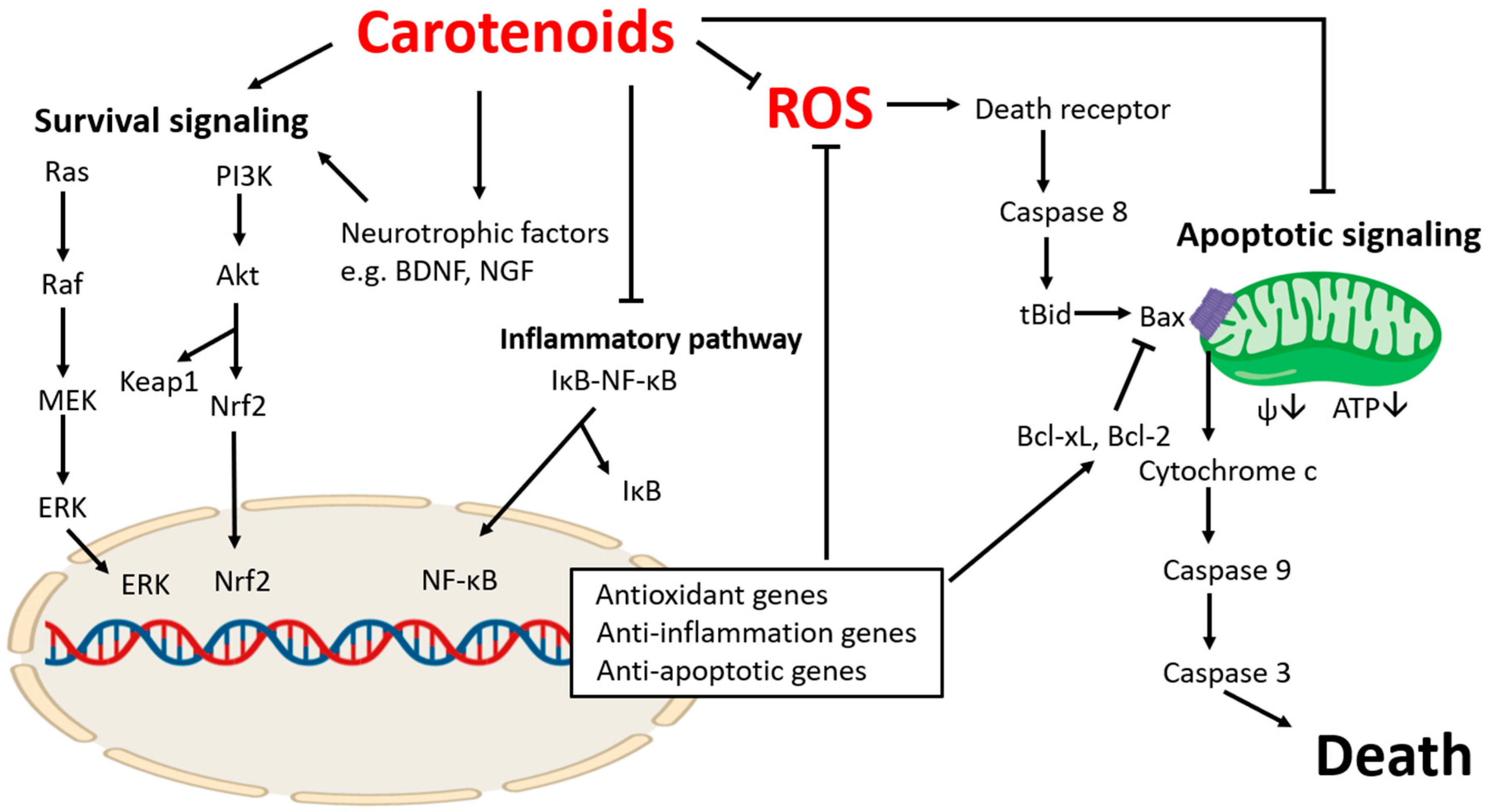
Table 1.
Current studies investigating roles of carotenoids in apoptotic pathways of brain related diseases.
Table 1.
Current studies investigating roles of carotenoids in apoptotic pathways of brain related diseases.
| Preventing Loss of Bcl-2 | Preventing Loss of Bcl-xL | Preventing Accumulation of Bax | Preventing Accumulation or Release of Cytochrome C | Preventing Accumulation or Activation of Caspase 3 | |
|---|---|---|---|---|---|
| β-carotene | [120] | [120] | [120] | ||
| Lycopene | [80,83,89] | [89] | [80,83,89] | [77,83,90] | [80,83] |
| Lutein | [172,173,174,193] | [173] | [173,174,193] | [174] | [173,193] |
| Astaxanthin | [140] | [136] | [137] | ||
| Zeaxanthin | [194] | ||||
| Fucoxanthin | [188] | [188] | [184] | [184,188] |
© 2020 by the authors. Licensee MDPI, Basel, Switzerland. This article is an open access article distributed under the terms and conditions of the Creative Commons Attribution (CC BY) license (http://creativecommons.org/licenses/by/4.0/).
Share and Cite
MDPI and ACS Style
Park, H.-A.; Hayden, M.M.; Bannerman, S.; Jansen, J.; Crowe-White, K.M. Anti-Apoptotic Effects of Carotenoids in Neurodegeneration. Molecules 2020, 25, 3453. https://doi.org/10.3390/molecules25153453
AMA Style
Park H-A, Hayden MM, Bannerman S, Jansen J, Crowe-White KM. Anti-Apoptotic Effects of Carotenoids in Neurodegeneration. Molecules. 2020; 25(15):3453. https://doi.org/10.3390/molecules25153453
Chicago/Turabian StylePark, Han-A, Mary Margaret Hayden, Sydni Bannerman, Joseph Jansen, and Kristi M. Crowe-White. 2020. "Anti-Apoptotic Effects of Carotenoids in Neurodegeneration" Molecules 25, no. 15: 3453. https://doi.org/10.3390/molecules25153453