Article

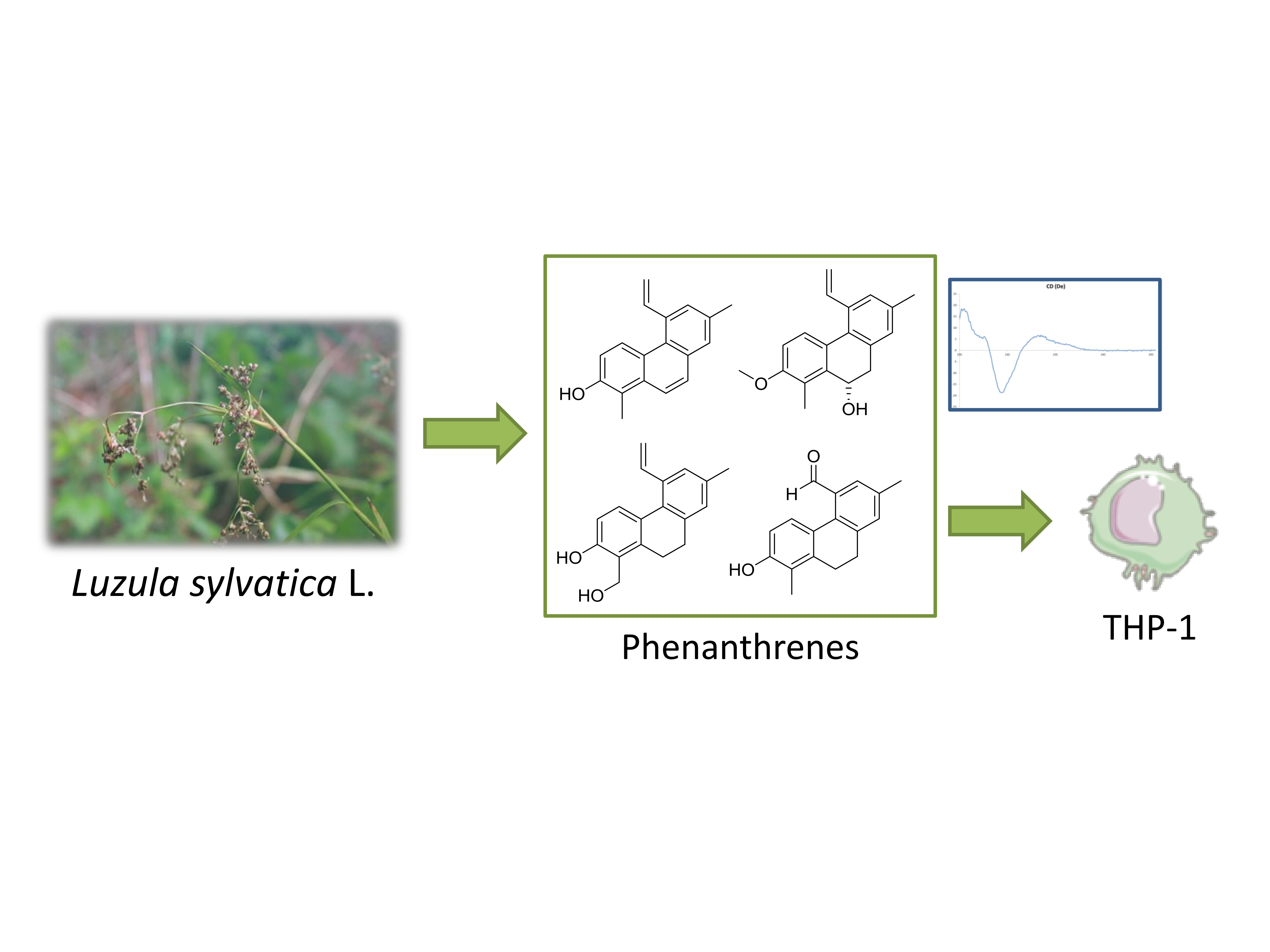
Anti-Inflammatory and Cytotoxic Potential of New Phenanthrenoids from Luzula sylvatica

by
Maël Gainche
1,
Isabelle Ripoche
1,*,
François Senejoux
2,*,
Juliette Cholet
2,
Clémence Ogeron
2,
Caroline Decombat
2,
Ombeline Danton
3,
Laetitia Delort
2,
Marjolaine Vareille-Delarbre
2,
Alexandre Berry
2,
Marion Vermerie
2,
Didier Fraisse
2,
Catherine Felgines
2,
Edwige Ranouille
4,
Jean-Yves Berthon
4,
Julien Priam
5,
Etienne Saunier
5,
Albert Tourrette
6,
Yves Troin
1,
Florence Caldefie-Chezet
2 and
Pierre Chalard
1
add Show full author list remove Hide full author list
1
Université Clermont-Auvergne, CNRS, SIGMA Clermont, ICCF, F-63000 Clermont-Ferrand, France
2
Université Clermont-Auvergne, INRA, UNH, Unité de Nutrition Humaine, CRNH Auvergne, F-63000 Clermont-Ferrand, France
3
Pharmaceutical Biology, Pharmacenter, University of Basel, Klingelbergstrasse 50, 4056 Basel, Switzerland
4
Greentech, Biopôle Clermont-Limagne, 63360 Saint-Beauzire, France
5
Dômes Pharma, 3 Rue André Citroën, 63430 Pont-du-Château, France
6
AltoPhyto, 7 rue des Gargailles, 63370 Lempdes, France
*
Authors to whom correspondence should be addressed.
Molecules 2020, 25(10), 2372; https://doi.org/10.3390/molecules25102372
Submission received: 21 April 2020 / Revised: 17 May 2020 / Accepted: 19 May 2020 / Published: 20 May 2020
(This article belongs to the Special Issue New Approaches for the Isolation of Natural Products)

Abstract

:
Phenanthrenoids have been widely described, in the Juncaceae family, for their biological properties such as antitumor, anxiolytic, anti-microbial, spasmolytic, and anti-inflammatory activities. The Juncaceae family is known to contain a large variety of phenanthrenoids possessing especially anti-inflammatory and cytotoxic properties. Luzula sylvatica, a Juncaceae species, is widely present in the Auvergne region of France, but has never been studied neither for its phytochemical profile nor for its biological properties. We investigated the phytochemical profile and evaluated the potential anti-inflammatory activities of L. sylvatica aerial parts extracts. A bioassay-guided fractionation was carried out to identify the most active fractions. Nine compounds were isolated, one coumarin 1 and eight phenanthrene derivatives (29), including four new compounds (4, 5, 8 and 9), from n-hexane and CH2Cl2, fractions. Their structures were established by HRESIMS, 1D and 2D NMR experiments. The biological properties, especially the anti-inflammatory/antioxidant activities (ROS production) and antiproliferative activity on THP-1, a monocytic leukemia cell line, of each compound, were evaluated. Three phenanthrene derivatives 4, 6, and 7 showed very promising antiproliferative activities.

Graphical Abstract

1. Introduction

Juncaceae represents a large family of plants with nearly 500 different species distributed in seven genera, the most representative ones are the Juncus and the Luzula genera (around 350 and 110 species respectively).
Plants belonging to this family are widespread all around the world, the Juncus and Luzula genera grow in both hemispheres, generally in badly drained soils. Several studies described the use of Juncaceae species for the traditional treatment of dysuria, fidgetiness, irritability, insomnia, and inflammation [1]. In China, J. inflexus is commonly used for its sedative effect, as J. effusus and J. conglomeratus are used for skin diseases [2,3] in the Basque region. Considering Luzula species, no ethnopharmacological uses have been reported yet. The biological activities of Juncaceae seem mostly due to the presence of flavonoids [4], coumarins [5], terpenoids [6], and phenolic acid derivatives [7]. In addition, phenanthrene derivatives exerting potent anti-inflammatory [8,9] and cytotoxic activities [10,11] have also been identified in several members of the family. Such constituents are scarcely distributed in the plant kingdom and have mainly been reported in the Orchidaceae [11] and the Combretacae families [12,13]. Regarding Juncaceae, most of the chemical studies focused on the Juncus genus leading to the identification of more than 100 phenanthrenoids [1]. Even if a large variety of phenanthrenoids have been described, most of them possess a phenanthrene or a 9,10-dihydrophenanthrene backbone, often substituted at positions 2, 5 and 7 with hydroxyl, methyl, or vinyl groups. A vinyl group at position 5 on the phenanthrene skeleton could be considered as a chemotaxonomic marker for Juncus species [11].
To our knowledge, very limited investigations have been reported on the Luzula species especially on Luzula sylvatica (L. sylvatica) [10]. This species is widely present in the Auvergne area, a region of France which possesses diversified flora, particularly due to the variety of landscapes and climatic conditions. L. sylvatica, commonly named Great Wood-Rush, is a perennial plant, measuring between 30 and 110 cm, and growing mostly in wet soils (humid forest, in the shade). The numerous dark green basal leaves, possessing small white cils, are between 6 and 12 mm wide and 10–25 cm long. The inflorescence is loose, subdivided in open panicles, and flowers are brownish, sessile and group by 2–5 in glomerules [14].
Like most of the Juncaceae plants, L. sylvatica seems to contain phenanthrenoids [1,15]. This class of compounds is known to different have biological properties such as antitumor, anxiolytic, antimicrobial, spasmolytic and anti-inflammatory activities [11]. Considering the strong chemotaxonomical and biological interests of phenanthrenoids, the present study aimed to investigate the chemical composition of a methanolic extract from L. sylvatica aerial parts focusing particularly on this chemical class. In addition, anti-inflammatory and antiproliferative properties of the studied extract and its isolated compounds were evaluated.

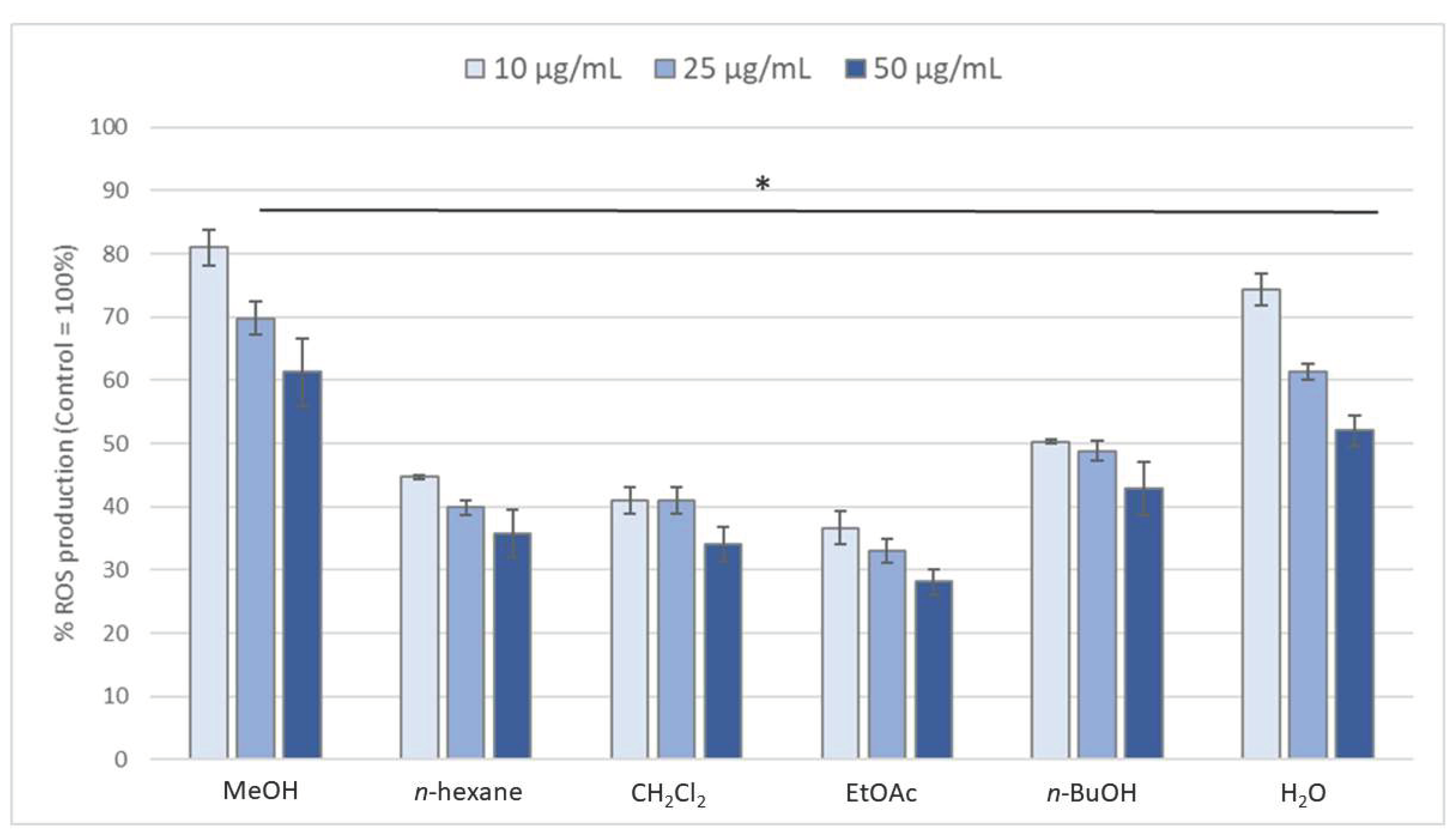
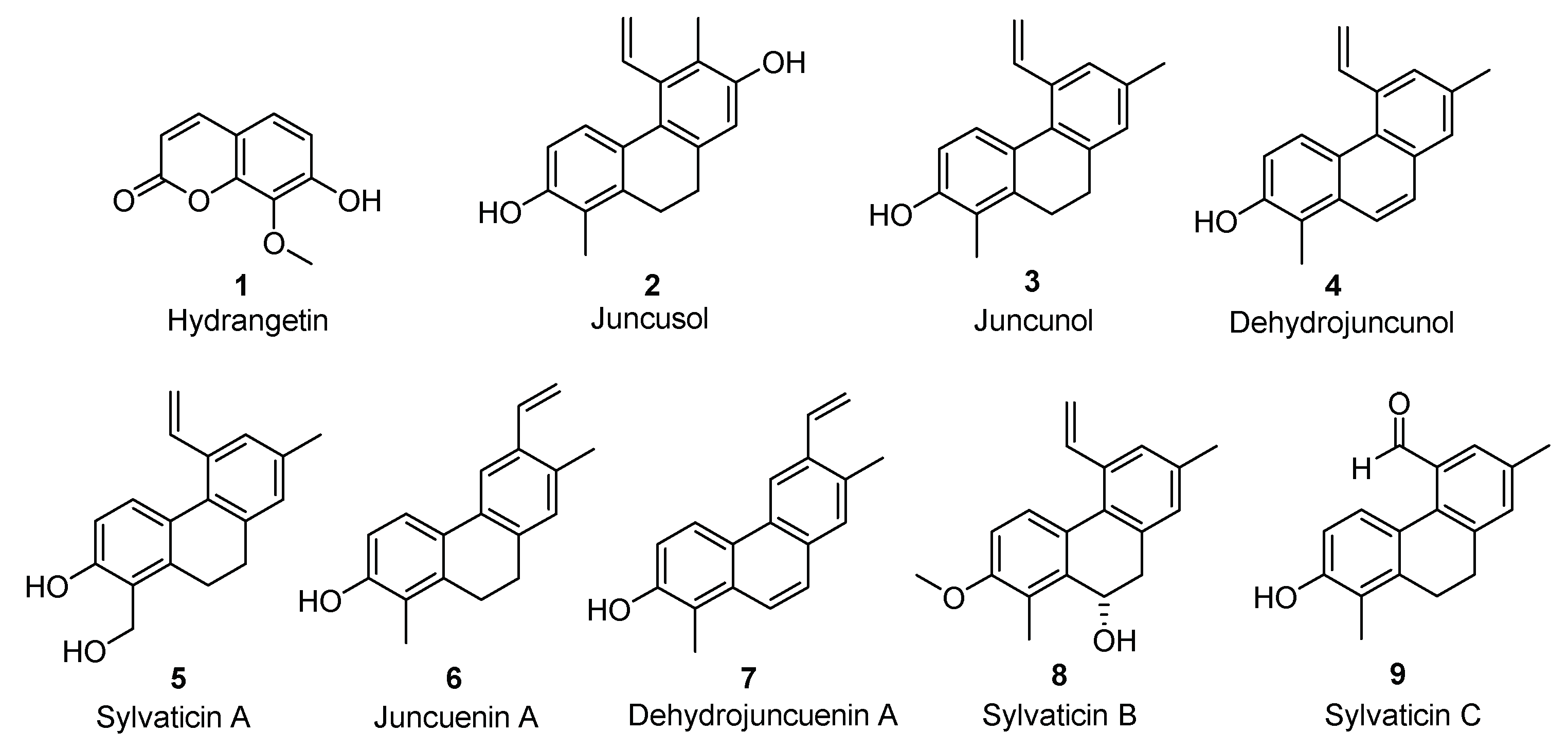
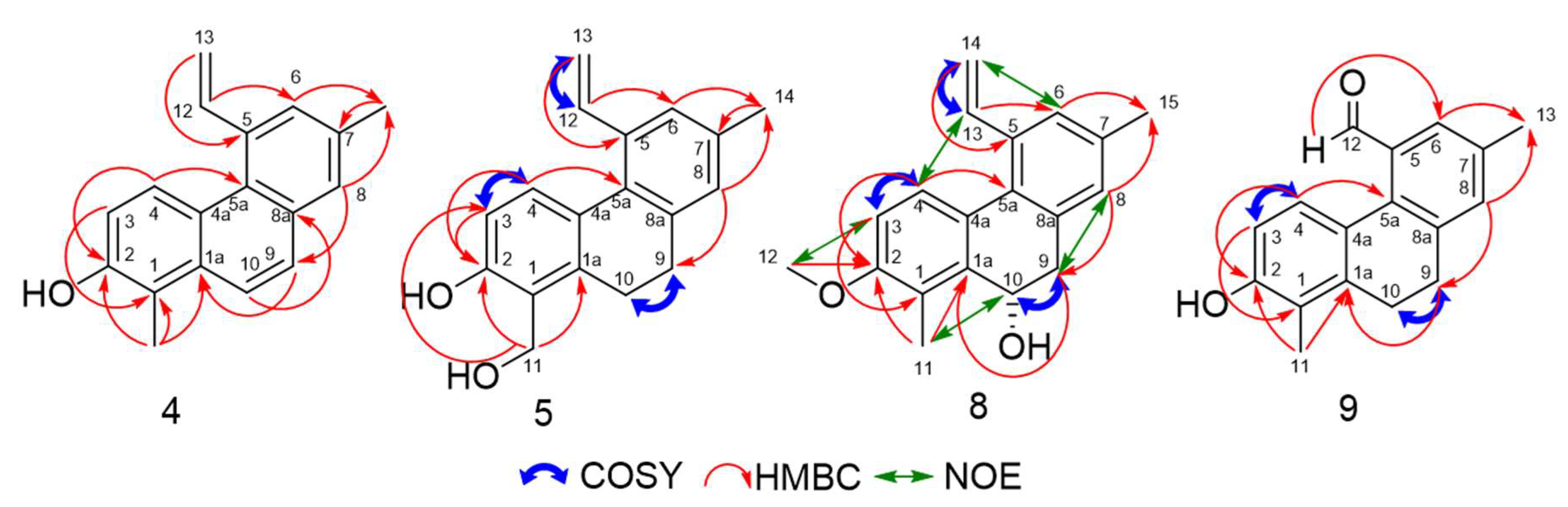
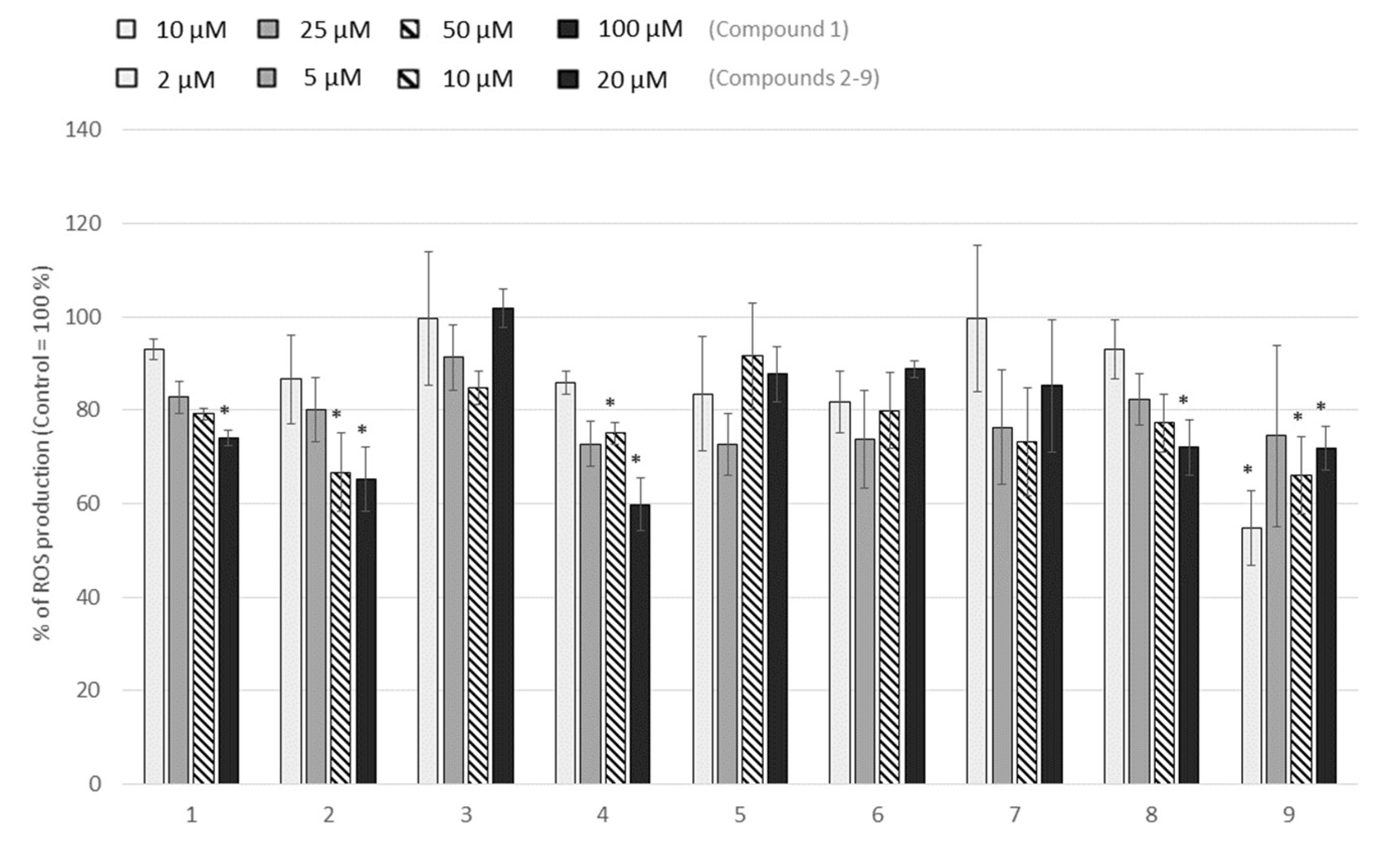
2. Results and Discussion

A methanolic extract, prepared from the aerial parts of L. sylvatica, was partitioned using increasing polarity liquid-liquid extraction. The five fractions ((n-hexane), dichloromethane (CH2Cl2), ethyl acetate (EtOAc), n-butanol (n-BuOH) and water) were tested for their anti-inflammatory/antioxidant activities by evaluating the production of ROS (reactive oxygen species) from blood leucocytes. All the fractions were able to decrease significantly the ROS production (Figure 1). The EtOAc, CH2Cl2 and n-Hex fractions were the most active ones (with a decrease of 60 to 75% compared to control). Analysis of the EtOAc fraction by LC-MS, LC-MS2, and UV spectrometry, in comparison with standards, led to the characterisation of luteolin, as its major constituent, as well as 1,3-O-dicaffeoylglycerol (ananasate) [16,17,18] and 3’,5,5’,7-tetrahydroxyflavanone [19]. Thus, the activity of EtOAc fraction could be due to the presence of luteolin, a flavone known for its anti-inflammatory properties [20,21].
Further purifications were performed on the n-hexane and the CH2Cl2 fractions which could potentially contain phenanthrene derivatives. From CH2Cl2 fraction, three compounds, namely juncusol (2) [1,22], compound 4 [23], and hydrangentin (1) [24,25], were isolated (Figure 2).
n-hexane fraction was first purified by steric exclusion method using biobeads, then by column chromatography. Six major compounds were isolated, juncunol (3) [23], juncuenin A (6), dehydrojuncuenin A (7) [26] and three new compounds (5, 8, and 9), with original phenanthrene structures, were identified.
The HRESIMS of compound 4, isolated as a pale-yellow oil, furnished deprotonated ion peak [M − H] at m/z 247.1127 (calcd 247.1128) corresponding to a molecular formula of C18H16O. The 1H NMR data showed the presence of three vinylic protons, six aromatic methine protons, two methyl groups and a hydroxyl group. A comparison of the NMR data of compound 4 with those obtained for juncunol and the HRESIMS data, suggested the presence of a phenanthrene backbone. The 13C NMR data confirm the presence of the vinylic function (δC 142.0 and 114.2 ppm) and the hydroxyl group (δC 151.1 ppm). HMBC correlations from H-13 (δH 5.78 and 5.44) to C-5 (δC 137.1) and from H-12 (δH 7.47) to C-6 (δC 130.6) suggested that the vinyl group was located at C-5. The HMBC correlations from H-6 (δH 7.45) and H-8 (δH 7.60) to C-14 (δC 21.3) suggested that the first methyl group was located at position 7. Moreover, a HMBC correlation from H-14 (δH 2.54) to C-7 (δC 134.9) confirmed this hypothesis. The HMBC correlation from H-8 (δH 7.60) to C-9 (δC 127.8) and the downshield chemical shifts of H-9 and H-10 protons (δH 7.69 and 7.89 ppm respectively) allowed us to confirm the skeleton of compound 4 as a phenanthrene backbone. The chemical shift of C-5a (δC 127.3) was determined based on the HMBC correlations from H-4 (δH 8.65), H-6 (δH 7.45), H-8 (δH 7.60) and H-9 (δH 7.69) to C-5a. The chemical shift of C-1a (δC 133.4) was confirmed by HMBC correlations from H-4 (δH 8.65) and H-9 (δH 7.69) to C-1a. The positions of the second methyl and the hydroxyl groups were determined by HMBC data. Correlations from H-11 (δH 2.61) to C-1 (δC 117.3), C-1a (δC 133.4) and C-2 (δC 151.1), from H-3 (δH 7.08) to C-1 (δC 117.3) and from H-4 (δH 8.65) to C-2 (δC 151.1) suggested that the second methyl group was located at C-1 and the hydroxyl group at C-2. The chemical shift of C-4a (δC 125.8) was confirmed by HMBC correlation from H-3 (δH 7.08) and H-10 (δH 7.89) to C-4a. The chemical shift of C-8a (δC 132.0) was confirmed by HMBC correlation from H-10 (δH 7.89) to C-8a. All these data confirmed the structure of compound 4 as 1,7-dimethyl-5-vinylphenanthrene-2-ol, named dehydrojuncunol.
Concerning compound 5, the HRESIMS furnished deprotonated ion peak [M − H] at m/z 265.1242 corresponding to a molecular formula of C18H18O2 (calculated 265.1234). Comparison with the 1H and the 13C NMR spectra of compound 3 suggested that compound 5 possesses a 9,10-dihydrophenanthrene backbone with a vinyl group located at C-5, a methyl group at C-7 and a hydroxyl group at C-2. The 1H and 13C data established the presence of a hydroxylmethyl group (δH 5.01), singlet for two protons (δC 60.2). HMBC correlations from H-11 (δH 5.01) and C-2 (δC 155.3), C-1a (δC 138.4) and C-3 (δC 113.4) and the correlation from H-3 (δH 6.78) and C-2 (δC 155.3), C-1a (δC 138.4) suggested that the hydroxyl group is located at C-2 and the hydroxyl methylene group at C-1. The structure of the new isolated compound 5 was confirmed as 1-hydroxymethyl-7-methyl-5-vinyl-9,10-hydrophenanthrene-2-ol, named sylvaticin A.
For compound 8, isolated as a pale-yellow oil, the molecular formula of C19H20O2 was established by HRESIMS (m/z 263.1426, [M + H − H2O]+; calculated m/z 263.1430) as well as the presence of a hydroxyl group. The chemical shift of a methine at δC 64.0 suggested the presence of the hydroxyl group on a non-aromatic carbon. 1H NMR data showed the presence of three vinylic protons, two methyl groups and a methoxy group. Comparison with the 1H and the 13C NMR spectra of compound 3 suggested that compound 8 is a 9,10-dihydrophenanthrene with a vinyl group located at C-5 and two methyl groups at C-1 and at C-7. The position of the methoxy group was determined by HMBC data. Correlations from H-4 (δH 7.56) and H-12 (δH 3.88) to C-2 (δC 157.1) suggested that the methoxy group is located at C-2. The structure of compound 8 was confirmed by NOESY. This experiment was performed in CD3OD in order to obtain two different chemical shifts for the protons of the two methyl groups (δH 2.33 and 2.35 respectively for H-11 and H-15) which had the same chemical shift in CDCl3 (δH 2.37). The data showed Overhauser effects between H-3 (δH 6.87) and H-12 (δH 3.88), H-4 (δH 7.56) and H-13 (δH 7.01), H-6 (δH 7.26) and H-14 (δH 5.72), H-8 (δH 7.09) and H-9 (δH 3.15), H-10 (δH 5.14) and H-11 (δH 2.37) as well as H-13 (δH 7.01) and H-14’(δH 5.28). The structure of the new compound 8 was determined as 2-methoxy-1,7-dimethyl-5-vinyl-9,10-dihydrophenanthrene-10-ol, named sylvaticin B. The CD spectrum of 8 showed a negative Cotton effect at 239 nm and a positive Cotton effect at 280 nm, in agreement with the (S) configuration of carbon 10 according to literature [27].
The HRESIMS of compound 9 furnished a deprotonated ion peak [M − H] at m/z 251.1077 (calcd 251.1078) corresponding to a molecular formula of C17H16O2. As the 1H and13C NMR data were strongly similar to compound 3 NMR data, we could identify compound 9 as a 9,10-dihydrophenanthrene backbone possessing three substituents, two methyl groups at C-1 and C-7, and a hydroxyl group at C-2. The 1H and 13C NMR data showed the presence an aldehyde function with the corresponding carbon at δC 193.5 ppm (CH) and a singlet proton at δH 10.07 ppm. As previously reported in literature [28,29,30], the aldehyde function was located on C-5. This was confirmed by the HMBC correlations from H-12 (δH 10.07) to C-6 (δC 127.2). The position of all the substituents and the chemical shifts of all the protons and the carbons were attributed using HMBC and COSY correlations as described on Figure 3. The structure of the new compound 9 was determined as 2-hydroxy-1,7-dimethyl-9,10-dihydrophenanthrene-5-carbaldehyde, named sylvaticin C.
The nine isolated compounds were tested on leucocytes ROS production as described above for the five fractions. Four of them inhibited ROS production significantly in a dose-dependent manner: compounds 2, 4, 8, and 1 (Figure 4), although the last one had to be tested at higher concentration to be effective.
In addition to their anti-inflammatory activity, phenanthrenes were known to exhibit promising in vitro antiproliferative activities on various cancer cell lines [9,31,32]. Thus, the cytotoxicity of the isolated compounds was evaluated with a resazurin assay on THP-1, a monocytic leukemia cell line.
Table 1 summarized the IC50 values obtained for each compound. With exception of compounds 1 and 9, all components revealed a strong cytotoxic activity, with IC50 below 15 µM. The most effective compounds were 4, 6, and 7 (IC50 of 3, 6, and 5 µM, respectively).

3. Materials and Methods

3.1. General Experimental Procedure

Nuclear magnetic resonance (NMR) spectra were recorded on an Avance III HD 400 MHz or 500 MHz (Bruker, Rheinstetten, Germany) spectrometer with CDCl3 as solvent (otherwise specified). All HPLC analyses were performed on an Agilent 1260 Infinity apparatus, with DAD detector (Agilent, Santa Clara, CA, USA) equipped with an Uptisphere C18-3 (250 × 4.6 mm, 5 µm) column (Interchim, Montluçon, France). LC-MS were carried out on an UHPLC Ultimate 3000 RSLC chain and an Orbitrap Q-Exactive (Thermo Scientific, Waltham, MA, USA) with the column mentioned above. Preparative chromatography was performed with a Varian prepstar Model SD-1 (Varian, Santa Clara, CA, USA) on an Uptisphere C18-3 (250 × 21.6 mm, 5 µm) semi-preparative column (Interchim). For all analyses, the mobile phase was a mixture of 0.1% (v:v) formic acid in water (phase A) and 0.1% (v:v) formic acid in acetonitrile (phase B). The gradient of phase A was 100% (0 min), 80% (10 min), 73% (35 min), 0% (40–50 min) and 100% (51–60 min). The flow rate was 0.8 mL/min, and the injection volume was 5 µL.
Flash chromatography was performed on a SPOT II FLASH (Interchim) with a monochromator detector. All chemical standards and references were purchased from Extrasynthese (Genay, France).
ECD spectra were recorded in methanol (200 μg/mL) on a Chirascan CD spectrometer using 1 mm path precision cells (110 QS, Hellma Analytics, Müllheim, Germany).

3.2. Plant Material

The aerial parts of L. sylvatica were collected in June 2017 (Cantal). A voucher specimen (CLF 110940) was deposited at the UniVegE herbarium of the Clermont Auvergne University.

3.3. Extraction and Isolation

Aerial parts of L. sylvatica (1.6 kg) were air-dried at room temperature in a dark room, powdered, then extracted with methanol (3 × 20 L each 24 h) and dried under vacuo to yield a greenish solid crude extract (195 g). This crude extract was then dissolved in water and extracted three times with increase solvent polarity to give five fractions: n-hexane (16.3%), CH2Cl2 (0.5%), EtOAc (2.0%) n-butanol (20.6%) and water (60.6%).
A part of the n-hexane fraction (12.11 g out of 21.87 g) was then partitioned with Biobeads SX-3 column chromatography (from Bio-Rad, Hercules, USA) in THF to afford 4 fractions (1–4). Fraction 4 (216.3 mg) was then partitioned through a silica gel column chromatography with cyclohexane - EtOAc as eluent (from 95:5 to 0:100, v:v) to afford six fractions (1 to 6). Fraction 1 contained only one major product: juncunol (26.3 mg) (3). Fraction 2 (77.2 mg) was subjected to semi-preparative chromatography with MeCN-H2O + 0.1% formic acid (75:25, v:v) to obtain juncuenin A (6) (4.9 mg) and dehydrojuncuenin A (7) (2 mg). Fraction 3 (29.6 mg) was purified by semi-preparative chromatography using MeCN-H2O + 0.1% formic acid (75:25, v:v) as eluent to obtain sylvaticin B (8) (3.1 mg) and sylvaticin C (9) (1.7 mg). Fraction 5 (36.7 mg) was purified with a silica gel chromatography column with cyclohexane-EtOAc (75:25, v:v) as eluent to afford sylvaticin A (5) (20.8 mg).
Part of the CH2Cl2 fraction (631 mg out of 983 mg) was subjected to a flash chromatography (normal phase) to afford 11 fractions (1 to 11). Fraction 2 was purified by silica gel column chromatography with cyclohexane-EtOAc (8:2 then 6:4, v:v) to afford juncusol (2) (4 mg). Fraction 3 (34.7 mg) was purified by silica gel column chromatography with cyclohexane-EtOAc (from 8:2 to 5:5, v:v) to obtain dehydrojuncunol 4 (3.4 mg). Fraction 5 was purified by silica gel chromatography column with cyclohexane-EtOAc-MeOH (from 6:4:0 to 0:9:1, v:v) to obtain hydrangetin (1) (25.3 mg).
1,7-Dimethyl-5-vinyl-phenanthrene-2-ol/ dehydrojuncunol (4). Pale yellow oil; 1H and 13C NMR data, Table 2, HRMS m/z 247.1127 [M − H] (calcd for C18H16O, 247.1128), more data in Supplementary Materials.
1-Hydroxymethyl-7-methyl-5-vinyl-9,10-hydrophenanthrene-2-ol/ sylvaticin A (5). Pale yellow oil; 1H and 13C NMR data, Table 2, HRMS m/z 265.1242 [M − H] (calcd for C18H18O2, 265.1234).
2-Methoxy-1,7-dimethyl-5-vinyl-9,10-dihydrophenanthrene-10-ol/ sylvaticin B (8). Pale yellow oil; 1H and 13C NMR data, Table 2, HRMS m/z 263.1426 [M + H − H2O]+ (calcd for C19H18O, 263.1430).
6-Hydroxy-2,5-dimethyl-9,10-dihydrophenanthrene-4-carbaldehyde/ sylvaticin C (9). Pale yellow oil; 1H and 13C NMR data, Table 2, HRMS m/z 251.1077 [M − H] (calcd for C17H16O2, 251.1078).

3.4. Blood Leucocytes ROS Production

Blood was collected from healthy human volunteers (n = 3–6; Etablissement Français du Sang, EFS, Clermont-Ferrand, France). Donors gave their written informed consent for the use of blood samples for research purposes under EFS contract n°16-21-62 (in accordance with the following articles L1222-1, L1222-8, L1243-4 and R1243-61 of the French Public Health Code). Blood leukocytes were obtained by hemolytic shock using ammonium chloride solution. Leucocytes were then washed with RPMI 1640 medium (GIBCO, ThermoFisher Scientific), centrifuged and suspended in supplemented RPMI (fetal bovine serum (FBS) 10%, gentamicin 50 μg/mL and glutamine (Gln) 2 mM). Cells were placed in 96-well plates (Cell Wells™, Corning, NY, USA), incubated with the extracts or the compounds and dihydrorhodamine 123 (DHR 123, 1 μM, Cayman Chemical Company, Ann Arbor, MI), and stimulated, or not, by 1 µM phorbol myristate acetate (PMA) for 60 min. The fluorescence intensity of rhodamine 123, which is the product of dihydrorhodamine 123 oxidation by ROS, was recorded (excitation/emission: 485/538 nm) using a microplate fluorometric reader (Tecan Spark®, Mämmedorf, Switzerland) [33,34].

3.5. Cytotoxicity Evaluation of the Compounds

The human monocytic leukemia cell line, THP-1 (American Type Culture Collection) was cultured and propagated at 37 °C in a humidified atmosphere of 5% CO2 in a RPMI 1640 medium supplemented with 10% FBS, gentamicin 50 μg/mL and Gln 2 mM. Cells were placed in 96-well plates and incubated with the compounds for 24 h. Then resazurin (25 µg/mL) was added to the medium to track their viability. Fluorescence (excitation/emission: 544/590 nm) was recorded for 2 h using a microplate fluorometric reader (Tecan Spark®) [35].

4. Conclusions

Phenanthrenoids are common secondary metabolites in plants of the Juncaceae family, possessing antiproliferative activities. From aerial parts of L. sylvatica, a Juncaceae species widely present in Auvergne region in France, we isolated nine compounds from the apolar n-hexane and CH2Cl2 fractions: coumarin 1 and eight phenanthrenoids (29). Among them, four new compounds were characterized: dehydrojuncunol 4, sylvaticin A 5, sylvaticin B 8, uncommonly substituted at C-10, and sylvaticin C 9. We demonstrated that phenanthrenoids 2, 4, and 8 inhibited ROS production significantly in a dose-dependent manner.
Compounds 4, 6, and 7 showed moderate cytotoxic effects on THP-1 cells with IC50 lower than 6 µM. Further investigations will be carried out on the most active compounds, especially on new phenanthrenoid 4 in order to study its antiproliferative activity on other cancer cells and to explore its mechanism of action.

Supplementary Materials

The NMR data are available online.

Author Contributions

Conceptualization, F.C.-C., P.C.; Formal analysis, O.D.; Funding acquisition, P.C.; Investigation, M.G., J.C., C.O.; Supervision, I.R., F.S., C.D., P.C.; Writing–original draft, M.G., I.R., F.S., P.C., J.C.; Writing–review & editing, M.G., I.R., F.S.; P.C., J.C., L.D., M.V.-D., A.B., M.V., D.F., C.F., Y.T., E.R., J.-Y.B., J.P., E.S., A.T. All authors have read and agreed to the published version of the manuscript.

Funding

This research was funded by the Regional Council of Auvergne Rhône Alpes (Conseil Régional d’Auvergne Rhône Alpes), the European Fund for Regional Economic Development (FEDER, AV0015282), Greentech and Dômes Pharma. We thank gratefully AltoPhyto for providing raw materials and Clermont Auvergne University Herbarium for voucher specimens and identification of plant material.

Acknowledgments

The authors thank Martin Leremboure for LC-MS and LC-MS2 analyses and Mounir Traika for NMR experiments.

Conflicts of Interest

The authors declare no conflict of interest.

References

  1. Bús, C.; Tóth, B.; Stefkó, D.; Hohmann, J.; Vasas, A. Family Juncaceae: Promising source of biologically active natural phenanthrenes. Phytoch. Rev. 2018, 17, 833–851. [Google Scholar] [CrossRef] [Green Version]
  2. Liao, Y.-J.; Zhai, H.-F.; Zhang, B.; Duan, T.-X.; Huang, J.-M. Anxiolytic and sedative effects of dehydroeffusol from Juncus effusin mice. Planta Med. 2010, 77, 416–420. [Google Scholar] [CrossRef] [PubMed]
  3. Menedez-Baceta, G.; Aceituno-Mata, L.; Molina, M.; Reyes-García, V.; Tardío, J.; Padro-de-Sanayana, M. Medicinal plants traditionally used in the northwest of the Basque Country (Biscay and Alava), Iberian Peninsula. J. Ethnoparmacol. 2014, 152, 113–134. [Google Scholar] [CrossRef]
  4. Li, H.-X.; Deng, T.-Z.; Chen, Y.; Feng, H.-J.; Yang, G.-Z. Isolation and identification of phenolic constituents from Juncus effusus. Acta Pharm. Sin. 2007, 42, 174–178. [Google Scholar]
  5. Della Greca, M.; Fiorentino, A.; Isidori, M.; Previtera, L.; Temussi, F.; Zarrelli, A. Benzocoumarins from the rhizomes of Juncus acutus. Tetrahedron 2003, 59, 4821–4825. [Google Scholar] [CrossRef]
  6. Shan, C.-Y.; Ye, Y.-H.; Jiang, H.-F.; Zhang, J. Study on chemical constituents isolated from Juncus effusus. J. Chin. Med. Mat. 2008, 31, 374–376. [Google Scholar]
  7. Dong-Zhea, J.; Zhi-Daa, M.; Geroge, C.Y.C.; Munekazu, I.; Toshiyuki, T. Two p-coumaryl glycerides from Juncus effusus. Phytochemistry 1996, 41, 545–547. [Google Scholar] [CrossRef]
  8. Behery, F.A.A.; Naeem, Z.E.M.; Maatooq, G.T.; Amer, M.M.A.; Wen, Z.H.; Sheu, J.H.; Ahmed, A.F. Phenanthrenoides from Juncus acutus L., new natural lipopolysaccharide-inducible nitric oxide synthase inhibitors. Chem. Pharm. Bull. 2007, 55, 1264–1266. [Google Scholar] [CrossRef] [Green Version]
  9. Ma, W.; Zhang, Y.; Ding, Y.-Y.; Liu, F.; Li, N. Cytotoxic and anti-inflammatory activities of phenanthrenes from the meduillae Juncus effusus L. Arch. Pharm. Res. 2016, 39, 154–160. [Google Scholar] [CrossRef]
  10. Kovács, A.; Vasas, A.; Hohmann, J. Natural phenanthrenes and their biological activity. Phytochemistry 2008, 69, 1084–1110. [Google Scholar] [CrossRef]
  11. Tóth, B.; Hohmann, J.; Vasas, A. Phenanthrenes: A promising group of plant secondary metabolites. J. Nat. Prod. 2018, 81, 661–678. [Google Scholar]
  12. Dawe, A.; Pierre, S.; Tsala, D.E.; Habtemariam, S. Phytochemical constituents of Combretum Loefl. (combretaceae). Pharm. Crops 2013, 4, 38–59. [Google Scholar] [CrossRef]
  13. Katerere, D.R.; Gray, A.I.; Nash, R.J.; Waigh, R.D. Phytochemical and antimicrobial investigations of stilbenoids and flavonoids isolated from three species of Combretaceae. Fitoterapia 2012, 83, 932–940. [Google Scholar] [CrossRef]
  14. Dumé, G.; Gauberville, C.; Mansion, D.; Rameau, J.-C. Flore Forestière Française 1; CNDF: Desmaures, QC, Canada, 2018; pp. 1774–1775. [Google Scholar]
  15. Tóth, B.; Chang, F.-R.; Tsong-Long, W.; Hwang, L.; Szappanos, Á.; Mándi, A.; Hunyati, A.; Tibor, K.; Jakab, G.; Hohmann, J.; et al. Screening of Luzula species native to the Carpathian Basin for anti-inflammatory activity and bioactivity-guided isolation of compounds from Luzula luzuloides (Lam.) Dandy & Wilmott. Fitoterapia 2017, 116, 131–138. [Google Scholar]
  16. Hu, J.; Lin, H.; Shen, J.; Lan, J.; Ma, C.; Zhao, Y.; Lei, F.; Xing, D.; Du, L. Developmental toxicity of orally administered pineapple leaf extract in rats. Food Chem. Toxicol. 2011, 49, 1455–1463. [Google Scholar] [CrossRef] [PubMed]
  17. Kang, J.; Price, W.E.; Ashton, J.; Tapsell, L.C.; Johnson, S. Identification and characterization of phenolic compounds in hydromethanolic extracts of sorghum wholegrains by LC-ESI-MSn. Food Chem. 2016, 211, 215–226. [Google Scholar] [CrossRef] [Green Version]
  18. Vargaa, M.; Jójárt, R.; Fónad, P.; Mihály, R.; Palágyi, A. Phenolic composition and antioxidant activity of colored oats. Food Chem. 2018, 268, 153–161. [Google Scholar] [CrossRef]
  19. Novakovic, M.; Djordjevic, I.; Todorovic, N.; Trifunovic, S.; Andjelkovic, B.; Mandic, B.; Jadranin, M.; Vuckovic, I.; Vajs, V.; Milosavljevic, S.; et al. New aurone epoxide and auronolignan from the heartwood of Cotinus coggygria Scop. Nat. Prod. Res. 2019, 33, 2837–2844. [Google Scholar] [CrossRef]
  20. Mizuno, M.; Yamashita, S.; Hashimoto, T. Enhancement of anti-inflammatory and anti-allergic activities with combination of luteolin and quercetin in in vitro co-culture system. Food Sci. Technol. Res. 2017, 23, 811–818. [Google Scholar] [CrossRef] [Green Version]
  21. Seelinger, G.; Merfort, I.; Schempp, C. Anti-oxydant, anti-inflammatory and anti-allergic activities of luteolin. Planta Med. 2008, 74, 1667–1677. [Google Scholar] [CrossRef]
  22. Tóth, B.; Liktor-Busa, E.; Kúsz, N.; Szappanos, Á.; Mándi, A.; Kurtán, T.; Urbán, E.; Hohmann, J.; Chang, F.-R.; Vasas, A. Phenantherenes from Juncus inflexus with antimicrobial activity against methicillin-resistant Staphylococcus aureus. J. Nat. Prod. 2016, 79, 2814–2823. [Google Scholar] [CrossRef] [PubMed]
  23. Abdel-Mogib, M. New 9,10-dihydrophenanthrenes derivatives from two Juncus species. Alex. J. Pharm. Sci. 2001, 15, 13–15. [Google Scholar]
  24. Dar, M.-Y.; Ara, T.; Akbar, S. A new prenylated coumarin from Daphne oleoides and its cytotoxic activity. Chem. Nat. Compd. 2019, 55, 5–7. [Google Scholar] [CrossRef]
  25. Liu, Z.-L.; Tian, X. The components of Cacalia tangutica. B. Kor. Chem. Soc. 2004, 25, 1078–1080. [Google Scholar]
  26. Wang, X.-Y.; Ke, C.-Q.; Tang, C.-P.; Yuan, D.; Ye, Y. 9,10-Dihydrophenathrenes and Phenanthrenes from Juncus setchuensis. J. Nat. Prod. 2009, 72, 1209–1212. [Google Scholar] [CrossRef]
  27. Resnick, S.M.; Gibson, D.T. Regio- and stereospecific oxidation of 9,10-dihydroanthracene and 9,10-dihydrophenanthrene by naphthalene dioxygenase: Structure and absolute stereochemistry of metabolites. Appl. Environ. Microbiol. 1996, 62, 3355–3359. [Google Scholar] [CrossRef] [Green Version]
  28. Della Greca, M.; Fiorentino, A.; Mangoni, L.; Molinaro, A.; Monaco, P.; Previtera, L. Cytotoxic 9,10-dihydrophenanthrenes from Juncus effusus L. Tetrahedron 1993, 49, 3425–3432. [Google Scholar] [CrossRef]
  29. Wang, Y.-G.; Wang, Y.-L.; Zhai, H.-F.; Liao, Y.-J.; Zhang, B.; Huang, J.-M. Phenanthrenes from Juncus effusus with anxiolytic and sedative activities. Nat. Prod. Res. 2012, 26, 1234–1239. [Google Scholar] [CrossRef]
  30. Su, X.-H.; Yuan, Z.-P.; Li, C.-Y.; Zhong, Y.-J.; Du, H.-J.; Wen, Y.-Y.; Li, Y.-F.; Liang, B. Phenanthrenes from Juncus effusus. Planta Med. 2013, 79, 10447–10452. [Google Scholar] [CrossRef]
  31. Ishiuchi, K.; Kosuge, Y.; Hamagami, H.; Ozaki, M.; Ishige, K.; Ito, Y.; Kitanaka, S. Chemical constituents isolated from Juncus effusus induce cytotoxicity in HT22 cells. J. Nat. Med. 2015, 69, 421–426. [Google Scholar] [CrossRef]
  32. Miles, D.H.; Bhattacharyya, J.; Mody, N.V.; Atwood, J.L.; Black, S.; Hedin, P.A. The structure of juncusol. A novel cytotoxic dihydrophenanthrene from the Estuarine marsh plant Juncus roemerianus. J. Am. Chem. Soc. 1977, 99, 618–620. [Google Scholar] [CrossRef] [PubMed]
  33. Caldefie-Chézet, F.; Fusillier, C.; Jarde, T.; Laroye, H.; Damez, M.; Vasson, M.-P.; Guillot, J. Potential anti-inflammatory effects of Melaleuca alternifolia essential oil on human peripheral blood leukocytes. Phytother. Res. 2006, 5, 364–370. [Google Scholar] [CrossRef] [PubMed]
  34. Cholet, J.; Decombat, C.; Vareille-Delarbre, M.; Gainche, M.; Berry, A.; Ogéron, C.; Ripoche, I.; Delort, L.; Vermerie, M.; Fraisse, D.; et al. Comparison of the anti-inflammatory and immunomodulatory mechanisms of two medicinal herbs: Meadowsweet (Filipendula ulmaria) and harpagophytum (Harpagophytum procumbens). Int. J. Plant Anim. Environ. Sci. 2019, 9, 145–163. [Google Scholar]
  35. Ramdani, L.-H.; Talhi, O.; Taibi, N.; Delort, L.; Decombat, C.; Silva, A.; Bachari, K.; Vasson, M.-P.; Caldefie-Chezet, F. Effects of Spiro-bisheterocycles on proliferation and apoptosis in human breast cancer cell lines. Anticancer Res. 2016, 36, 6399–6408. [Google Scholar] [CrossRef] [Green Version]
Sample Availability: Samples of the compounds are not available from the authors.
Figure 1. ROS production of blood leucocytes, incubated with the methanolic extract or fractions (10, 25 and 50 µg/mL) and stimulated with PMA (1 µM) for 1 h. Values are expressed as percentage of the control (cells incubated with PMA and without extract). Data are shown as means ± SEM (n = 3–6); * p < 0.05 compared with Control. All results are expressed as a percentage, with control (i.e., cells with PMA but without extract) normalized as 100%.
Molecules 25 02372 g001
Figure 2. Isolated compounds from CH2Cl2 and n-hexane fractions.
Molecules 25 02372 g002
Figure 3. Key COSY, HMBC and NOESY correlations for compounds 4, 5, 8 and 9.
Molecules 25 02372 g003
Figure 4. ROS production of blood leucocytes, incubated with the compounds (2, 5, 10 and 20 µM for compounds 29 and 10, 25, 50 and 100 µM for compound 1) and stimulated with PMA (1 µM) for 1 h. Values are expressed as percentage of the control (cells incubated with PMA and without extract). Data are shown as means ± SEM (n = 3); * p < 0.05 compared with Control. All results are expressed as a percentage, with control (i.e., cells with PMA but without extract) normalized as 100%.
Molecules 25 02372 g004
Table 1. Cytotoxic effect of compounds 19 in THP-1 cells reported as IC50 in µM. Cells metabolic activity was determined after 24 h of incubation with or without compounds at different concentrations (2, 5, 10 and 20 µM for compounds 29 and 10, 25, 50 and 100 µM for compound 1) by resazurin assay (n = 3). Doxorubicine was used as a positive control at 1 µM for our experiments. At 1 µM, the viability of THP-1 decreased at around 80% (21.4 ± 3% of the fluorescence after 24 h of incubation, with negative control normalized as 100%).
CompoundIC50 (µM)
1>100
210
313
43
511
66
75
810
9>20
Table 2. 1H and 13C data for compounds 4, 5, 8 and 9 (in CDCl3).
Compound 4Compound 5Compound 8Compound 9
PositionδC typeδH (J in Hz)δC typeδH (J in Hz)δC typeδH (J in Hz)δC typeδH (J in Hz)
1117.3, C 126.8, C 124.1, C 121.7, C
2151.1, C 155.3, C 157.1, C 154.5, C
3114.8, CH7.08, d (9.1)113.5, CH6.78, d (8.5)109.2, CH6.87, d (8.4)112.7, CH6.76, d (8.4)
4127.4 CH8.65, d (9.1)130.6, CH7.49, d (8.5)128.5, CH7.56, d (8.4)129.4, CH6.92, d (8.4)
5137.1 C 135.1, C 135.4 C 140.1, C
6130.6, CH7.45, s127.4, CH7.25, s128.3, CH7.26, s127.2, CH7.64, s
7134.9, C 136.0, C 136.7, C 136.3, C
8128.2, CH7.60, s127.6, CH7.00, s130.1, CH7.09, s133.1, CH7.30, s
9127.8, CH7.69, d (9.1)30.0, CH22.69, m38.2, CH23.15, dd (16, 2.8)29.1, CH22.78, m
9’ 2.95, dd (16, 3)
10122.8, CH7.89, d (9.1)25.1, CH22.69, m64.0, CH5.14, m25.5, CH22.82, m
1111.3, CH32.61, s60.2, CH25.01, s11.2, CH32.37, s11.8, CH32.30, s
12142.0, CH7.47, dd (17.3, 10.7)138.8, CH6.93, dd (17.4, 10.8)55.8, CH33.88, s193.6, CH10.07, s
13114.2, CH25.78, dd (17.3, 1.7)114.1, CH25.70, dd (17.4, 1.3)139.1, CH7.01, dd (17, 10)21.1, CH32.41, s
13’ 5.44, dd (10.7, 1.7) 5.25 dd (10.8, 1.3)
1421.3, CH32.54, s21.2, CH32.37, s114.3, CH25.72, dd (17, 2)
14’ 5.28, dd (10, 2)
15 21.1, CH32.37, s
1a133.4, C 138.4, C 138.2, C 139.5, C
4a125.8, C 121.6, C 125.4, C 124.4, C
5a127.3, C 131.1, C 129.9, C 136.7, C
8a132.0, C 138.5, C 132.9, C 133.0, C
OH 4.94 brs 5.12, brs

