Article

Genome-Wide Transcriptome and Metabolome Analyses Provide Novel Insights and Suggest a Sex-Specific Response to Heat Stress in Pigs

1
Amimal Genomics and Bioinformatics Division, National Institute of Animal Science, RDA, Wanju 55365, Korea
2
Animal Nutrition and Physiology Team, National Institute of Animal Science, RDA, Wanju 55365, Korea
*
Author to whom correspondence should be addressed.
These authors contributed equally.
Genes 2020, 11(5), 540; https://doi.org/10.3390/genes11050540
Submission received: 5 March 2020 / Revised: 6 May 2020 / Accepted: 6 May 2020 / Published: 11 May 2020
(This article belongs to the Special Issue Pig Genomics and Genetics)

Abstract

:
Heat stress (HS) negatively impacts pig production and swine health. Therefore, to understand the genetic and metabolic responses of pigs to HS, we used RNA-Seq and high resolution magic angle spinning (HR-MAS) NMR analyses to compare the transcriptomes and metabolomes of Duroc pigs (n = 6, 3 barrows and 3 gilts) exposed to heat stress (33 °C and 60% RH) with a control group (25 °C and 60% RH). HS resulted in the differential expression of 552 (236 up, 316 down) and 879 (540 up, 339 down) genes and significant enrichment of 30 and 31 plasma metabolites in female and male pigs, respectively. Apoptosis, response to heat, Toll-like receptor signaling and oxidative stress were enriched among the up-regulated genes, while negative regulation of the immune response, ATP synthesis and the ribosomal pathway were enriched among down-regulated genes. Twelve and ten metabolic pathways were found to be enriched (among them, four metabolic pathways, including arginine and proline metabolism, and three metabolic pathways, including pantothenate and CoA biosynthesis), overlapping between the transcriptome and metabolome analyses in the female and male group respectively. The limited overlap between pathways enriched with differentially expressed genes and enriched plasma metabolites between the sexes suggests a sex-specific response to HS in pigs.

1. Introduction

Heat stress affects animal husbandry worldwide and is a major environmental factor that affects animal health and production [1,2]. The increasing environmental temperature due to global warming will particularly affect pigs due to their lack of functional sweat glands, which would have helped in endogenous heat dissipation [3,4]. Added to this, the thick subcutaneous adipose tissue in the pig, impedes effective radiant heat loss [5]. Moreover the increase in metabolic rate due to rapid lean tissue accretion increases endogenous heat production, exacerbating the innate inability of porcine animals to tolerate heat [3,6,7,8,9]. The estimated annual economic loss due to heat stress for the swine industry in the US alone is nearly US $300 to $450 million [5,10,11], mainly due to increased mortality and morbidity, altered carcass composition, decreased feed efficiency, inconsistent growth, reduced fecundity and poor sow performance [6], so heat stress in a pressing issue for worldwide pig production.
Quiniou and Noblet [12] have shown that the growth rate is normally between 18 and 25 °C and that the thermoregulatory response in pigs is activated at about 25 °C. Animals respond to heat stress by regulating physiological and metabolic changes, such as redistribution of blood flow from the body core to the periphery, and by reducing feed intake [13] to reduce metabolic heat production [14]. This has a significant effect on animal health and productivity [3]. The heat stress mitigation options available at present include expensive heat abatement processes, such as spray or floor cooling and nutritional supplementation strategies. Since the response to heat stress varies within a population, due to genetic variations, selection for thermal tolerance could be a better alternative [1,15]. However traditional breeding programs have limitations due to the difficulties in collecting precise records of thermal states of individual animals. To overcome this, production traits that are correlated with heat stress are used for selecting animals with higher thermal tolerance [16]. They, however, mask the actual effects of heat stress; therefore, a precise and efficient method for identifying genetic tolerance to heat stress is necessary. Identifying genetic and metabolic markers that are sensitive to heat stress will be the most important step for managing heat stress in pigs. These markers could also be putative candidates for identifying genomic variants for improving heat tolerance in pigs.
Previous studies have shown that heat stress triggers a complex array of gene expression and metabolic changes in livestock, resulting in the modulation of a wide variety of pathways involved in the heat stress response, the inflammatory response, DNA damage repair, chaperone use, etc. [5,17,18,19]. Similarly, a variety of metabolites involved in carbohydrate, amino acid, fatty acid and amine metabolism are modulated, including glucose, lactate, alanine, cysteine, isoleucine, etc. [20,21,22,23]. However, these studies did not involve integrated transcriptome and metabolome profiling, which could lead to better understanding of the effect of heat stress on swine physiology, gene expression and metabolism, which will help in designing and implementing innovative strategies to limit the economic losses due to heat stress on pig farm profitability. Therefore, in this study, we exposed 3-month-old pigs to heat stress and performed transcriptome and metabolome analysis using RNA-Seq and high resolution magic angle spinning (HR-MAS) NMR (nuclear magnetic resonance) to understand the gene expression and metabolome perturbations in response to heat stress.

2. Materials and Methods

2.1. Animals

The experiments were performed following the ethical guidelines laid down by the Institutional Animal Care and Use Committee (IACUC) of the National Institute of Animal Science (NIAS). The experimental procedures were reviewed and approved by IACUC of NIAS (NO 2017-1070). A total of 6 (3 barrows and 3 gilts) unrelated, 3-month-old Duroc pigs, weighing 56 ± 2.62 Kg, were used in this study. Animals were fed a corn and soybean meal based diet (Table 1), containing 18% crude protein, 4.9% crude fat, 4.6% crude fiber and 4.4% crude ash; the whole diet was formulated to provide 3450 Kcal/Kg. The animals had ad libitum access to feed and water. Water and feed intake were measured (Table S1). One-way analysis of variance (ANOVA) was performed in R to test for significance. Differences were considered to be significant at p < 0.05. Other than the typical heat stress responses, no significant differences in behavior were noted.

2.2. Heat Stress Experimental Setup

The heat stress (HS) experiment was carried out in an environmental chamber which had controls for temperature and humidity in NIAS. After acclimation to the chamber for 3 days, the experiments were performed. The animals were first kept at 25 °C and 60% relative humidity for 24 h, and at the end, 10 mL of blood was drawn via venipuncture and 5 mL of blood was stored in a Tempus Blood RNA tube (Life Technlogies, Carlsbad, CA, USA) for RNAseq analysis and the rest of the blood was stored in a vacutainer tube containing anticoagulants (EDTAK2), for the metabolomic analysis. The same animals were then exposed to 33 °C temperature and 60% relative humidity for 24 h and blood was drawn and stored in the same way.

2.3. RNA Isolation and Sequencing

The RNA was isolated using TRIzol Reagent (Invitrogen, Carlsbad, CA, USA) following the manufacturer’s guidelines. The integrity and quality of the RNA was assessed using 2100 Bioanalyzer and RNA 6000 Nano LabChip Kit (Agilent Technologies, Palo Alto, CA, USA). Only samples with RIN values >8 were used for library construction. The concentration of RNA was determined using NanoDrop ND-1000 spectrophotometer (NanoDrop Technologies, Wilmington, DE, USA). The sequencing library was constructed using Illumina TruSeq RNA sample preparation kit (Illumina, San Diego, CA, USA) following processes previously described [16]. The sequencing was performed on an Illumina HiSeq 2000 sequencer. The raw reads are available for download from sequence read archive (SRA), NCBI under the accession number SUB7043048.

2.4. Sample Preperation and Metabolite Analysis with 1H-NMR

36 μL of Plasma was mixed with 4 μL of D2O (20 mM TSP-d4), and the sample was then transferred to a 4 mm NMR nanotub, and 1H-NMR spectra were obtained by high-resolution magic-angle-spinning (HR-MAS) NMR spectra using an Agilent 600 MHz NMR spectrometer (Agilent Technologies, Palo Alto, CA, USA) with a 4 mm gHX Nanoprobe. A Carr–Purcell–Meiboom–Gill (CPMG) pulse sequence was used to reduce signals generated by macromolecules and water. The 1H-NMR spectra were measured as described previously [24]. The TSP-d4 peak at 0.0 ppm was used as a reference for calibrating chemical shifts.

2.5. Data Analysis

2.5.1. RNA-Seq Data

The processing of the data was previously described [25]. Briefly, after checking the quality of the raw reads using FastQC (version 0.11.5) [26] and trimming the adaptors and low-quality bases using TRIMMOMATIC (version 0.36) [27], the reads were aligned to the Pig reference genome (Sus scrofa 11.1) using HiSAT2 (version 2.05) [28]. The aligned reads were then counted using FeatureCounts (version 1.5.0) [29]. After correcting for batch and unknown effects using Svaseq [30], differential expression analysis was performed using DESeq2 [31]. Significant genes (FDR < 0.1) were identified, and functional enrichment analysis based on Gene Ontology (GO) under Biological Process and Molecular Function was performed with DAVID [32]. KEGG (Kyoto Encylopedia of Genes and Genomes (KEGG) pathway enrichment analysis was performed using ClueGO plugin [33] in cytoscape version 3.7.2 [34]. Goseq [35] was used for metabolic pathway enrichment analysis.

2.5.2. Metabolome Data

The metabolite quantification was performed using Chenomx NMR suite 7.1 (Chenomx, Edmonton, Canada). The generated spectra were binned with a binning size of 0.001 ppm, and normalized to the total area, and binned data were aligned with the icoshift algorithm of MATLAB R2013b (MathWorks, Natick, MA, USA). Principle component analysis (PCA) and statistical analyses were performed using MetaboAnalyst 4.0 [36]. Only features that were detected in at least 50% of samples were used.

2.5.3. Pathway Enrichment Analysis of Differentially-Enriched Metabolites

Significantly differentially-enriched metabolites (p < 0.05) were subjected to KEGG pathway analyses using the Pathway analysis module in MetaboAnalyst 4.0 [36]. A combination of quantitative enrichment and topology analysis using only curated metabolic pathways from the KEGG database was used in the analyses.

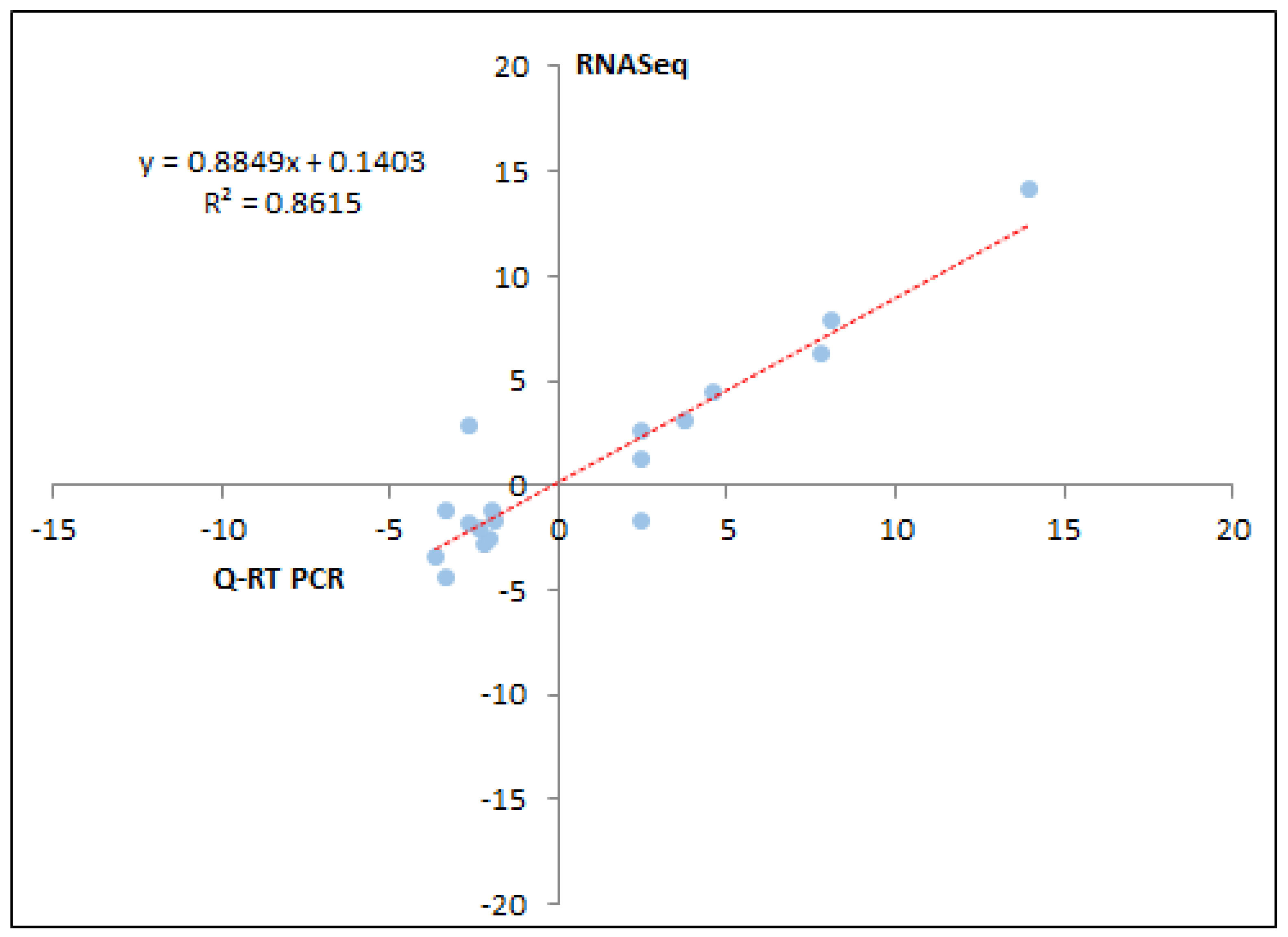
2.6. Real-Time PCR Validation

Real time reverse transcriptase PCR (qRT-PCR) was performed using gene-specific primes (Supplementary File S1). The PCR was performed on an ABI 7500 Real Time PCR system using Fast SYBR green master mix (Applied Biosystems, Foster City, CA, USA). A total of 18 genes (9 each from male and female differentially expressed gene (DEG) set) were analyzed. β-actin and GADPH (Glyceraldehyde-3-phosphate dehydrogenase) were used as endogenous controls. The stability of expression for each of those two genes was checked using GeNORM (https://genorm.cmgg.be/) against the same concentration of RNA from different samples. β-actin was the most stable and was used for normalizing the expression data of the target genes.

3. Results

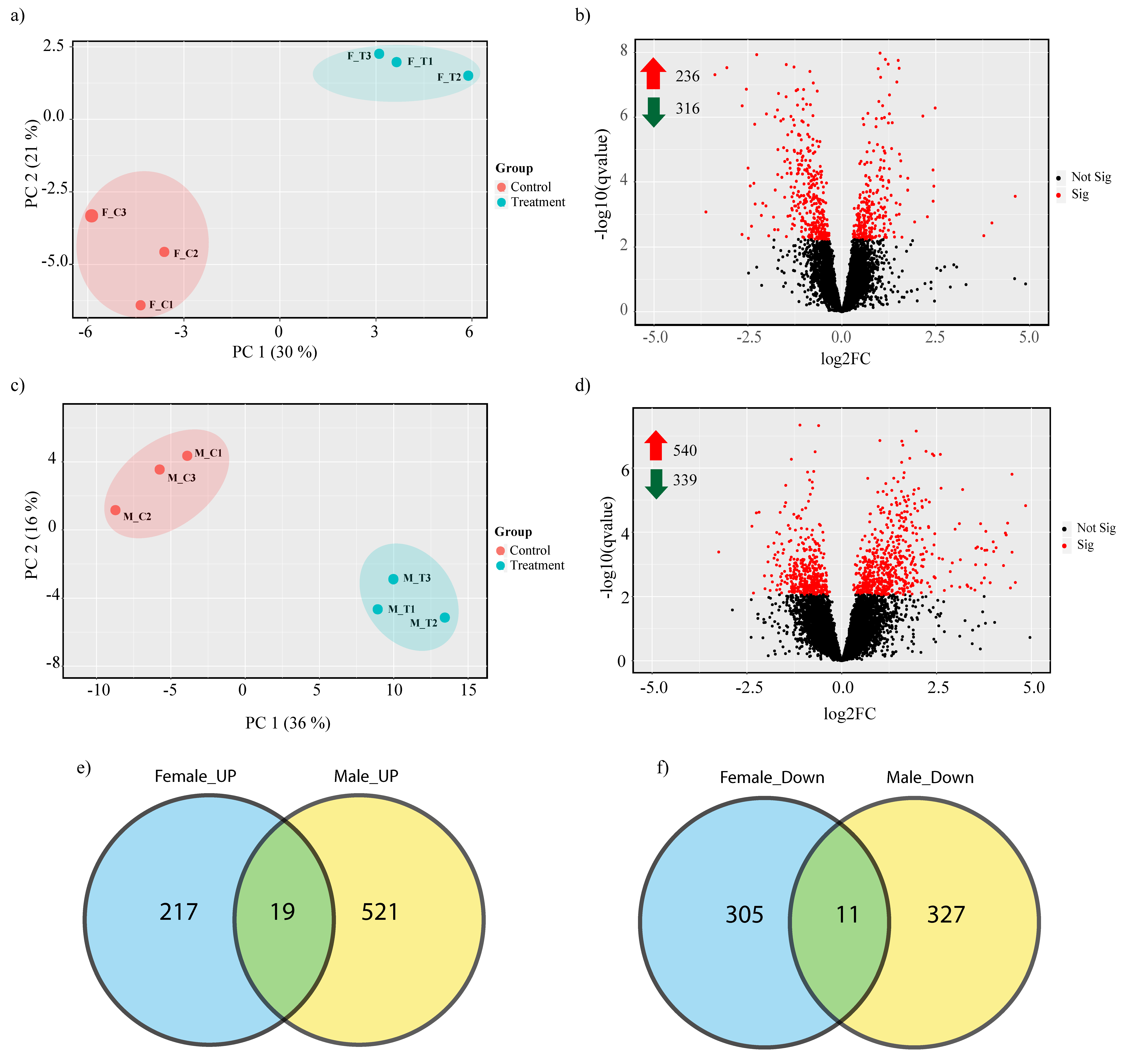
3.1. Transcriptome Alignment, Mapping and Principle Component Analysis

We investigated the impact of heat stress on Duroc pigs using high throughput RNA-Seq analysis. A total of 423.3 million 100 bp Paired-End (PE) reads corresponding to an average of 35.2 million reads per individual was generated. After trimming for adapters and low-quality reads, 410.5 million reads remained. The reads were mapped to the Sus scrofa reference genome at an average alignment rate of 96.8% (File S2). The reads were mapped to a total of 19,283 genes. Principal component analysis (PCA) (Figure S1) suggested that sex had a large effect on transcriptome difference between the groups, so we decided to compare the heat stress effect on male and female pigs separately. PCA showed that 30% and 36% of the expression variation was due to heat stress in female and male pigs respectively (Figure 1a,c); however, considerable within group variation was also observed, confirming previous reports that the heat stress response varies within populations due to underlying genomic variation [1,15].

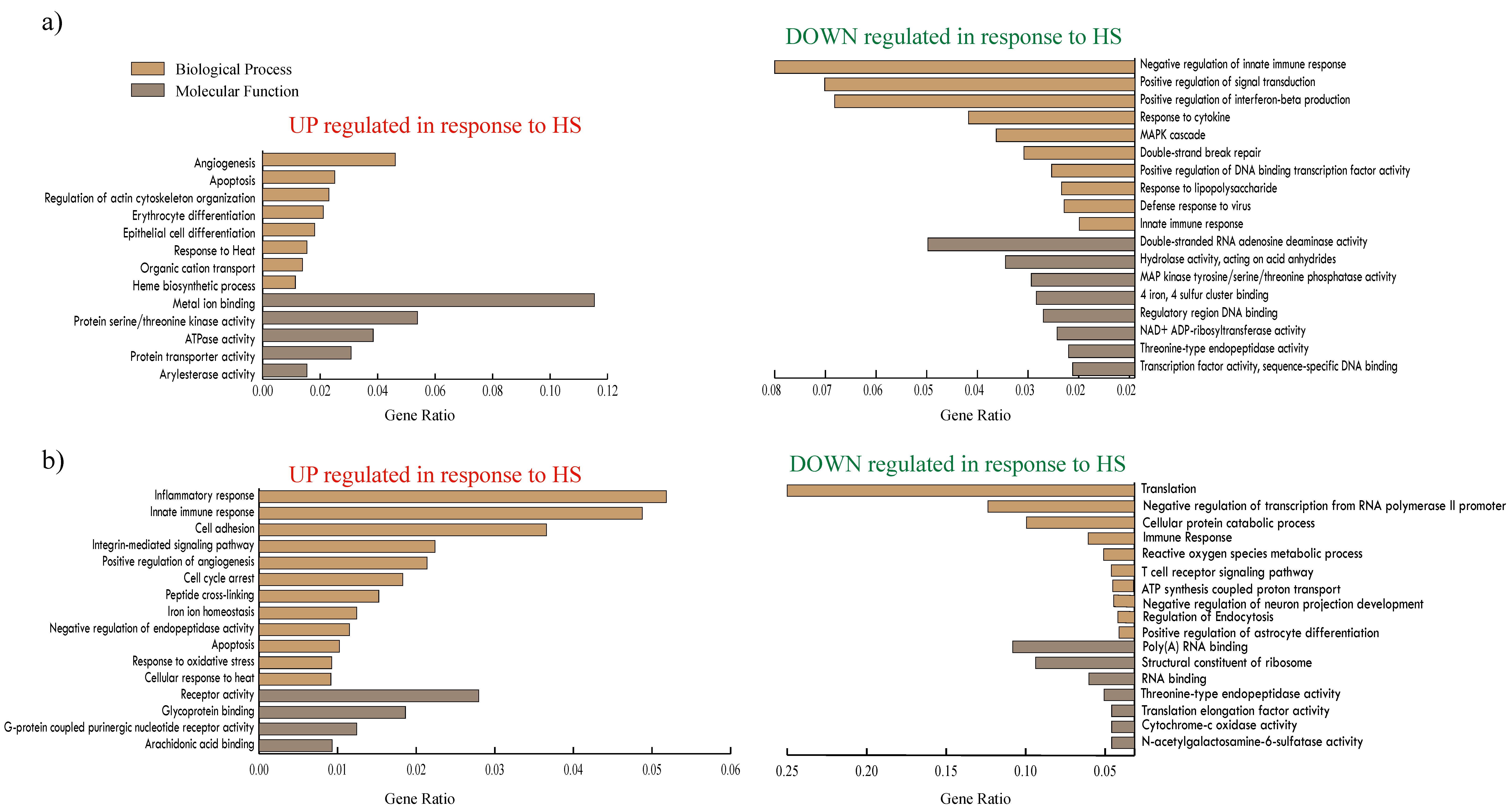
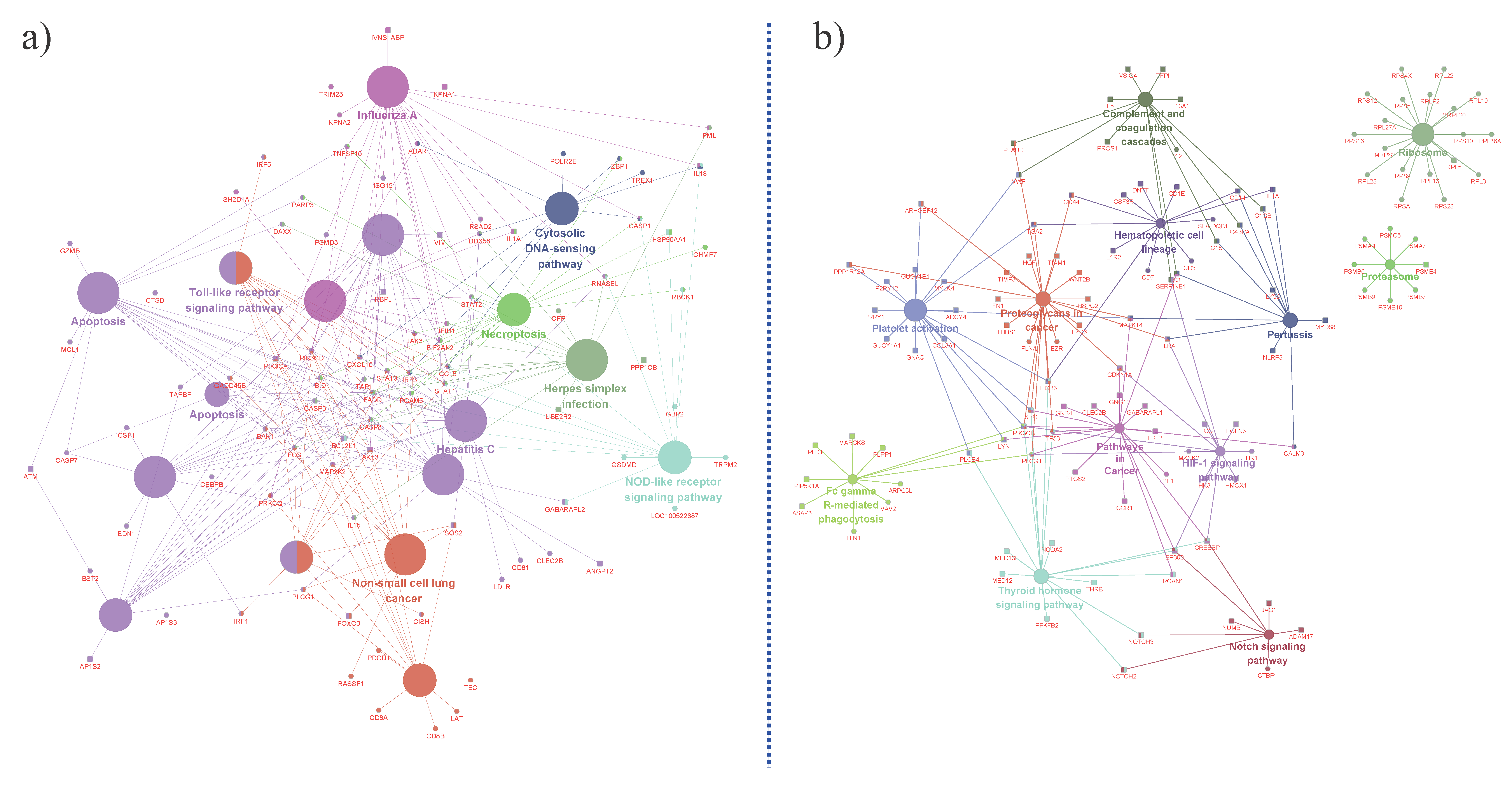
3.2. Porcine Transcriptome Response to Heat Stress

Heat stress resulted in 552 and 879 genes being significantly (FDR < 0.1) differentially expressed in male and female groups respectively. Out of those, 236 and 540 genes were up-regulated and 316 and 339 genes were down-regulated in female and male pigs respectively (Figure 1b,d, File S3). The genes modulated in response to heat stress were considerably different between the male and female groups, with only 19 up-regulated and 11 down-regulated genes being common between the groups (Figure 1e,f). Among the top up-regulated DEGs in the female group were HIST2H2AA4 (histone cluster 2H2A family member a4), MYH4 (myosin heavy chain 4), ABCA6 (ATP binding cassette subfamily A member 6), HSF4 (heat shock factor 4) and IL18 (interleukin 18), while top down-regulated DEGs included CXCL10 (C-X-C motif chemokine 10), IDO1 (indoleamine 2,3-dioxygenase 1), HES4 (Hes family BHLH transcription factor 4) and IFI6 (γ-interferon-inducible protein Ifi-16); in the male group the up-regulated genes included DES (Desmin), RAG1 (recombination activating 1), LSAMP (limbic system associated membrane protein), CD1E (CD1e molecule), HSPG2 (heparin sulfate proteoglycan 2) and down-regulated genes included CEBPE(CCAAT/enhancer binding protein), ZNF316 (Zinc Finger Protein 316), PXDC1(PX domain containing 1) and COX4I1 (cytochrome c oxidase subunit 4l 1).
Gene Ontology (GO) enrichment analysis of the significantly DEGs (FDR < 0.1) in the female group (Figure 2a) showed that among the up-regulated DEGs, “angiogenesis,” “apoptosis,” “response to heat,” “ heme biosynthetic process” and “ATPase activity” were enriched, whereas “negative regulation of innate immune response,” “positive regulation of signal transduction,” “response to cytokine,” “MAPK cascade,” “response to lipopolysaccharide,” “threonine-type endopeptidase activity,” etc., were enriched amongst the down-regulated genes. KEGG (Kyoto Encyclopedia of Genes and Genomes) pathway enrichment analysis of the female group DEGs (Figure 3a) showed that “apoptosis,” “non-small cell lung cancer,” “Toll-like receptor signaling pathway,” “necroptosis,” “NOD-like receptor signaling pathway” and “Influenza A” pathways were enriched.
In the male group, heat stress resulted in the up-regulation of genes functioning in “inflammatory response,” “innate immune response,” “cell adhesion,” “integrin-mediated signaling pathway,” “cell cycle arrest,” “positive regulation of angiogenesis,” “apoptosis,” “response to oxidative stress,” “cellular response to heat” and “glycoprotein binding.” Genes functioning in the “cellular protein catabolic process,” “regulation of endocytosis,” “negative regulation of astrocyte differentiation,” “cytochrome-c oxidase activity” and “N-acetylgalactosamine-6-sulfatase activity” were down-regulated (Figure 2b). Among KEGG pathways (Figure 3b), genes part of “complement and coagulation cascades,” “platelet activation,” “fc γ r-mediated phagocytosis,” “thyroid hormone signaling,” “pathways in cancer,” “proteoglycans in cancer,” and “Notch signaling” pathways were enriched. The top 10 DEGs from the female and male group were validated using q-RT PR analysis (quantitative Real-time PCR) (Figure 4); the correlation between RNA-Seq and q-RT PCR analyses was 0.861.

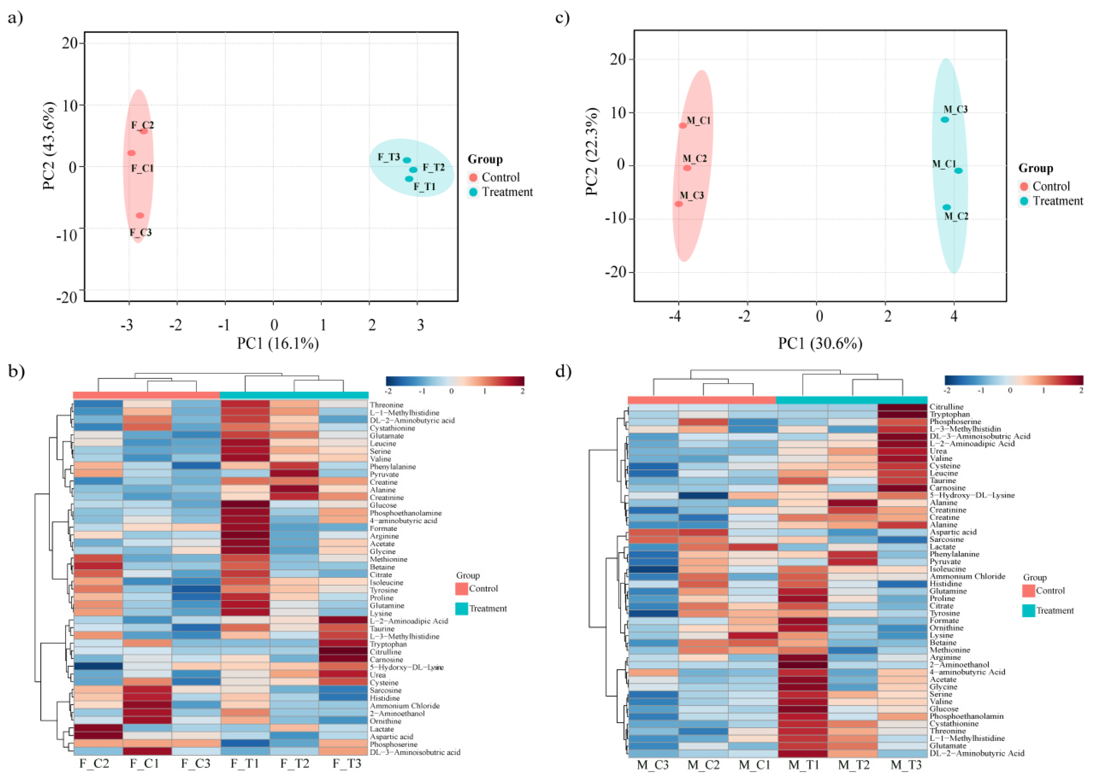
3.3. Porcine Metabolome Response to Heat Stress

We detected and identified a total of 46 metabolites (Figure 5b,d, File S4). After normalizing the sample (quantile normalization) and scaling the data (auto scaling) in MetaboAnalyst 4.0 [36], PCA and a heatmap were generated. The overall metabolic profiles of the groups resolved well in PC1 (Figure 5a,c), and hierarchical clustering based on metabolite concentration showed that the control and heat stress group were well separated. However, PC2 showed that there was considerable within-group variation in both male and female pigs. The fold change was calculated as a ratio between the two groups’ means. A Wilcoxon rank-sum test was performed, and 30 and 31 metabolites were found to significantly differ (p < 0.05) between the control and heat stressed groups in female and male groups respectively (File S4).
In the female group, out of the 30 metabolites that significantly differed between the heat stress and control groups, the concentrations of 21 metabolites increased while nine metabolites decreased in concentration. The concentrations of tryptophan, arginine, pyruvate, alanine, acetate and lactate were higher, while DL-plus allo-δ-hydroxylysine, creatine, urea, 3-aminoisobutric acid, 4-aminobutyric acid and 3-methylhistidine were lower in the heat stress group (File S4). In the male group samples, 18 of the 31 significantly differing metabolites increased, among which where aspartic acid, pyruvate, betaine, creatine and lactate, while 13 metabolites, including tryptophan, carnosine, valine, alanine and creatine, were significantly reduced in the heat stress group compared to the control group (File S4).

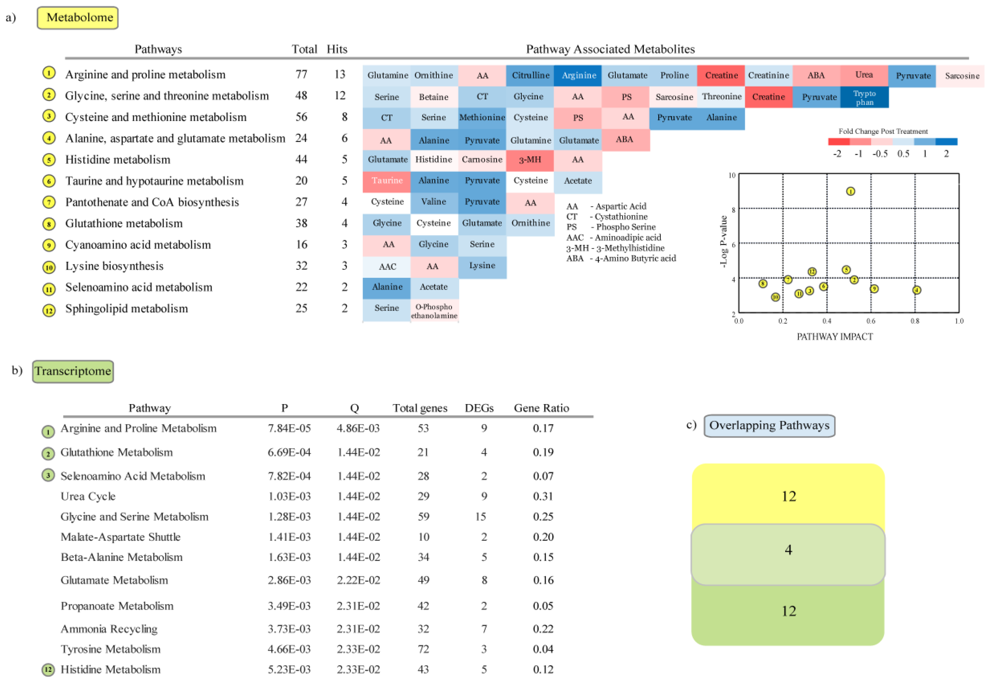
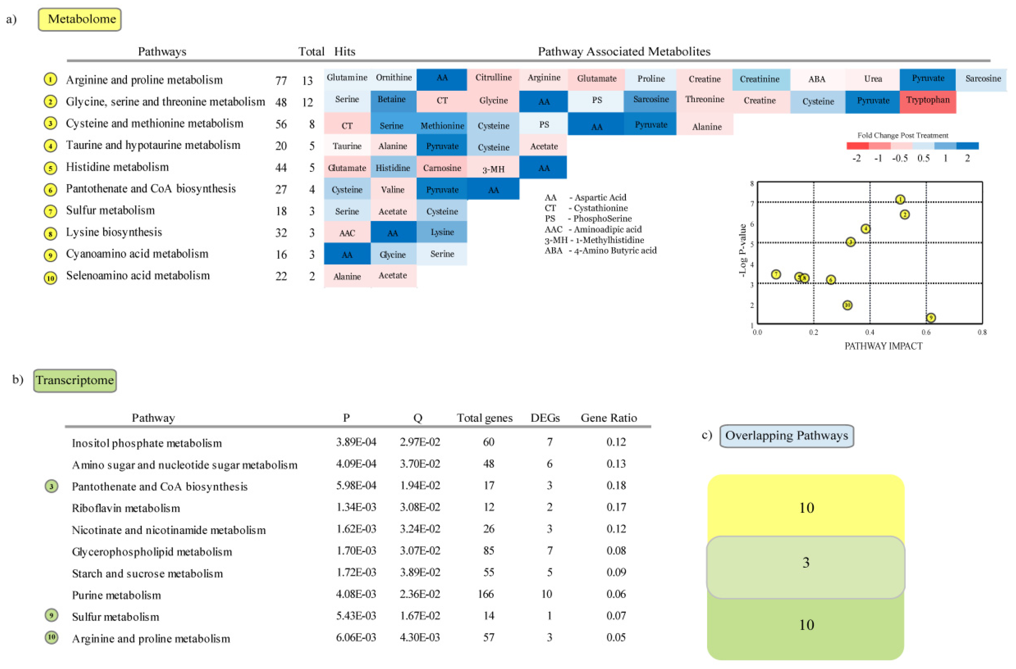
3.4. Metabolic Pathways Enriched in Response to Heat Stress

Pathway analysis was performed with metabolites that significantly differed between the heat stress and control group using the pathway enrichment module in MetaboAnalyst 4.0 [36]. Twelve pathways were significantly (p < 0.05) enriched in the female group (Figure 6a), including “arginine and proline metabolism,” “glycine, serine and threonine metabolism,” “cysteine and methionine metabolism,” “alanine, aspartate and glutamate metabolism” and “histidine metabolism”. We used goseq [35] to identify metabolic pathways in KEGG database that were significantly altered in the transcriptome, in response to heat stress. Among the twelve pathways significantly enriched (Figure 6b), four pathways were found to be commonly enriched with DEGs and DEMs (differentially enriched metabolites) (Figure 6c); these were “arginine and proline metabolism,” “glutathione metabolism,” “selenoamino acid metabolism” and “histidine metabolism.” Out of these, the first three pathways have been found to be enriched in response to oxidative stress induced by chronic HS in the pig [37,38,39].
Among the male animals, 10 metabolic pathways were enriched with differently enriched metabolites, including “arginine and proline metabolism,” “glycine, serine and threonine metabolism,” “cysteine and methionine metabolism,” “taurine and hypotaurine metabolism” and “histidine metabolism” (Figure 7a). The metabolic pathways enriched with DEGs (Figure 7b) included “inositol phosphate metabolism,” “amino sugar and nucleotide sugar metabolism,” “pantothenate and COA biosynthesis” and “riboflavin metabolism.” Comparative analysis of the metabolic pathways enriched with DEMs and DEGs showed that three metabolic pathways were common (Figure 7c); those were “pantothenate and CoA biosynthesis,” “sulfur metabolism” and “arginine proline metabolism.” Pantothenate and CoA biosynthesis was previously found to be enriched in response to HS in the pig [5,20].

4. Discussion

Heat stress occurs when the rate of heat accumulation exceeds the rate of heat loss; i.e., when the animal is removed from the thermal comfort zone. HS causes the core body temperature of a pig to rise [40]. Temperature increase due to global warming will be detrimental to swine health and production. The global temperature has already been raising at an average of 0.13 °C over the past 50 years [41]. Pigs are particularly vulnerable due to their lack of functional sweat glands, and the thick layer of subcutaneous adipose tissue that they possess which impedes effective radiant heat loss [42]. Heat stress triggers an adaptive stress response by regulating a complex array of genes and metabolites [16,18,21]. In this study we found significant differences in water and feed intake post heat stress, and a significant difference in water intake post heat stress treatment between male and female pigs (Table S1).
Several studies have suggested that gender might have an effect on the heat stress response in pigs [40,43,44,45]. The sex-specific response to heat stress could be due to the difference in surface area to mass ratio or due to body compositional difference (lean meat to fat mass) which affects radiant heat loss [43]. Barrows are reported to generally have greater backfat thickness than gilts [46]. Though barrows consume more feed and gain body weight more rapidly, gilts deposit proportionally more muscle and less fat in their carcass [43,47]. This sex-specific difference in body composition could significantly alter the heat stress response, as it affects the ability of the animal to maintain the core body temperature through radiant heat loss. Moreover, several hormones, such as prostaglandins [48], endogenous opioids [48] and glucocorticoids have roles in the thermoregulatory mechanism [49]. Cortisol, a glucocorticoid, is the mostly widely used biomarker for detecting stress [50,51]. The concentration of cortisol is 15% higher in barrows than in gilts [52]. HS-induced increases in the concentrations of catecholamines and glucocorticoids have been observed in many species [53,54]; the effect of HS on immune cells is suggested to be dependent upon these hormones [55,56,57]. Similarly, Rudolph et al. [40], found that oxidative stress did not result in muscle injury in barrows, while previous studies in gilts reported that oxidative stress causes muscle injury [58,59,60,61]. This led them to suggest that muscle response to heat stress could be partially dependent upon sex of the pig, and that muscle tissues in males could be more resistant to heat stress than in females. The result of our study concurs with all these reports; our study shows that HS in pigs, as evidenced from transcriptome expression and metabolome concentration, is sexually dimorphic.

4.1. Transcriptome Regulation in Response to Heat Stress

Transcriptome analysis showed that heat stress caused a profound shift in gene expression in the male group: 879 genes were found to be significantly differentially regulated in the male group, while 552 genes were DEs (differentially expressed) in the female group. Among the DEGs were several genes involved in Heat shock response. Heat shock proteins (HSPs) are expressed upon encountering stress [62,63]. Heat is proteotoxic and causes proteins to denature [64], HSPs act as chaperones in assisting in protein folding, thereby helping in avoiding protein aggregation [65] and maintaining cellular homeostasis [66]. HSPA5 (heat shock 70 kDa protein 5), HSP90AA1 (heat shock protein 90 α family class a member 1) and HSF4 (heat shock transcription factor 4) were up-regulated, in both male and female groups, while FKBP5 (FKBP prolyl isomerase 5) was up-regulated only in the male group. HSPA5 is a member of the HSP70 (heat shock protein 70) family of genes; it serves as an endoplasmic reticulum chaperone and as a sensor of protein misfolding [67]. HSP90AA1 a member of the HSP90 family takes part in several cellular functions such as regulating protein activity, transport and also in activating different signaling pathways by forming complexes with steroid receptors [68]. HSP90AA1 is essential for normal spermatogenesis in pigs [69], and regulation of HSP90AA1 protects cells from heat shock [70]. FKBP5 a member of immunophilin family, is a glucocorticoid receptor (GR)-regulating co-chaperone of heat shock protein 90, their expression are regulated by progestins and glucocorticoids [71], and are significantly correlated with plasma cortisol concentration in pig [72,73], suggesting that the male pigs were under considerable stress.
Several co-chaperones and genes involved in cellular response to heat stress were also DEs; these included HMOX1 (heme oxygenase 1), TRPM2 (transient receptor potential melastatin 2), which were up-regulated in the male group, IL1A (interleukin 1 α); up-regulated in both male and female groups, SOD1 (superoxide dismutase 1) down-regulated in male group and TFEC (Transcription factor EC), DAXX (death domain associated protein), and TRPM2 which were down-regulated in the female group. HMOX1 is an oxidative stress marker [74], and is up-regulated in response to oxidative stress [75]. SOD1 catalyzes the removal of superoxide radicals that are generated due to biological oxidation [76]. SOD expression was found to be down-regulated in porcine skeletal muscle in response to long-term (3 days) heat stress [58]. In this study SOD expression was down-regulated in the male group following heat stress; this along with the increased expression of HMOX1 suggests that the male group animals may have experienced significant oxidative stress following heat stress. Several genes involved in response to oxidative stress were up-regulated in the male group, including TRPM2 [77], LRRK2 [78], MAPK14 [79] and VNN1 [80]. In the female group PRDX2 [81], SLC7A11 [82] and FOXO3 [83] which protects cells from ROX were up-regulated, while TREX1 involved in cellular response to oxidative stress [84] and SETX (Senataxin) involved in defense against DNA damage due to oxidative stress [85] were down-regulated in the female group.
Immune response is closely linked with response to heat stress. Immune stimulation could occur either due to the hyper thermic effect on immune cells or due to indirect effects such as the activation of heat shock factors, which are potent immune modulators and can stimulate both innate and adaptive immune response [86]. Heat stress triggered a massive innate immune response in the male group, with several genes including CD14, BMX, S100A8, TLR4, MYD88, SRC, TLR2, SLEC5A, S100A9, PTK2B and VNN1 being up-regulated; interestingly in the female group, 13 genes involved in innate immunity were down-regulated; those included, TRIM5, ISG20, RSAD2, DDX58, PML, TRIM25, TEC, IRF3, TRIM26, SH2D1A, ANXA1, FADD and JAK3. In the male group, among the DEGs several inflammatory response genes were up-regulated; these included CD14, CCR1, TSPAN2, TLR4, THBS1, PTGER3, MYD88, C5AR2, SELP, TLR2 and CHI3L1, while inflammatory response was not enriched amongst the DEGs in the female group. Anti-tumor immune response were also markedly enhanced due to heat stress, genes involved in proteoglycans in cancer, choline metabolism in cancer, pathways in cancer were up-regulated in both male and female groups, while apoptotic genes were only triggered in the female group; these included genes PIK3CA, BCL2L1, AKT3 and ATM. Several studies have shown that heat stress triggers anti-tumor and apoptotic pathways possibly due to protein aggregation due to heat stress induced denaturation [16].

4.2. Metabolome Regulation in Response to Heat Stress

Metabolites are building blocks for growth and development; they are also key regulators and markers of animal health [87]. Blood metabolites are highly sensitive to environmental stress [88] and HS alters protein metabolism in a number of species [19,89,90,91]. Metabolome analysis showed that several plasma metabolites were enriched in response to HS in both male and female groups, this included, creatinine, histidine, lysine, methionine, ornithine, serine, proline and pyruvate, while 4-aminobutryic acid, Creatine, Taurine and Urea concentration was lower post HS. Plasma creatinine concentration has been found to be increased due to heat stress in several species including cattle, pigs and sheep [92,93,94]. Creatinine along with methylhistidine is a known indicator of muscle breakdown [95], the concentration of 1-Methylhistidine was also significantly increased in the female group. This indicates that the animals might be experiencing tissue break-down due to heat stress, the reason for such breakdown is not clear, however it could probably be due to increased protein catabolism, which is required for utilizing the carbon in amino acids for gluconeogenesis [96]. Methionine is a sulfur containing amino acid, and along with creatinine is involved in cellular antioxidant mechanism and is found abundantly on the surface of proteins exposed to very high oxidant fluxes [97,98]. The concentration of Pyruvate, an anti-oxidant [99] has been found to be increased under hyperthermia in pigs [100]. Pyruvate is also the precursor to alanine via alanine aminotransferase, and the entry of pyruvate into the TCA cycle through the pyruvate dehydrogenase complex is impaired due to heat stress, possibly due to the inactivation of PDH (Pyruvate dehydrogenase complex) complex due to the HS induced oxidative chain reactions generated by intracellular ROS (reactive oxygen species) [101]. Along with increased plasma concentration of Pyruvate due to the inactivation of PDH, the increased expression of PDK4 (Pyruvate Dehydrogenase Kinase 4) is suggested to serve as a mechanism to reduce substrate oxidation and mitochondrial ROS production to protect HS induced cellular damage [6]. The expression of PDK4 was significantly increased in the male group. Glycine stimulates protein synthesis, and has been found to inhibit oxidative stress in pig small intestine, the plasma concentration of glycine in the female group increased post HS while it reduced in the male group [102]. Similarly Alanine and Citrulline have protective effect against oxidative damage [103]. The difference in the plasma concentration of several of the above discussed metabolites together with increased expression of PDK4, HMOX1 and down-regulation of SOD1 indicates that the male group could have experience heat stress induced oxidative stress.
Several plasma metabolites were also oppositely enriched in the male and female groups. The plasma concentration of Tryptophan, 1-Methylhistidine, Acetate, Glycine, Aminoadipic acid, Alanine, Arginine, Citrulline, Cystathionine, Glutamate, Threonine, and Valine were all increased in the female group, while their concentration was reduced in the male group. Tryptophan metabolites are key neurotransmitters, that regulate immune response, increased plasma concentration of tryptophan could indicate intracellular protein degradation, and were found to increase the expression of apoptosis initiators in pig [104,105]. The plasma concentration of metabolites like glutamate, cystathionine and threonine are all associated with apoptosis and autophagy [106,107,108]. Several anti and pro apoptotic genes were also amongst the DEGs. This along with previously known effects of HS on protein denaturation and the proteotoxic effect of HS [16], and the increased concentration of amino acids that are by products of protein degradation suggest that HS induced cellular damage must have occurred in female group.

5. Conclusions

In conclusion, heat stress triggered a dynamic response in pigs; however, the response to heat stress was sex-specific. The reason for this sexual dimorphic response is not clear; however, evidence from other studies suggests that it could be due to the anatomical, physiological and hormonal differences. Future transcriptome and metabolome studies, along with blood parameters and hormonal analyses could provide more understanding about the effect of sex on the heat stress response in pigs. The results of this study along with increasing our understanding of porcine heat stress response will serve as a good reference for future studies. However, there are some limitations in this study, such as only six animals being used in this study for both transcriptome and metabolome analyses, and since only NMR was used for metabolite quantification, it resulted in limited detection of metabolites (only 48 metabolites) resulting in only a limited understanding of the effect of HS on metabolite accumulation, so complementing NMR analysis with mass spectrometric analysis might result in a more comprehensive understanding of the effect of HS on metabolite accumulation.

Supplementary Materials

The following are available online at https://www.mdpi.com/2073-4425/11/5/540/s1. File S1: List of primers used in the q-RT PCR analysis. File S2: Summary of the mapping and alignment statistics. File S3: List of DEGs identified in response to heat stress. File S4: List of metabolites found to be differentially enriched in response to heat stress. Figure S1: PCA using RNA-Seq and metabolome data from all samples. Table S1: Summary of the measurements of average daily feed and water intake.

Author Contributions

Conceptualization, K.S., S.D.L., G.-W.J. and J.-E.P.; methodology, K.S., S.Y.J., K.H.K., Y.K.L., M.K., Y.C.B. and S.D.L.; software, K.S.; validation, H.K. (Hana Kim); formal analysis, K.S., Y.C.B. and M.K.; investigation, K.S. and H.K. (Himansu Kumar); resources, J.-E.P. and S.D.L.; data curation, B.-H.C., G.-W.J. and S.Y.J.; writing—original draft preparation, K.S.; writing—review and editing, S.D.L. and J.-E.P.; visualization, K.S.; supervision, G.-W.J. and J.-E.P.; project administration, S.D.L.; funding acquisition, S.D.L. and J.-E.P. All authors have read and agreed to the published version of the manuscript.

Funding

This work was carried out with the support of the Cooperative Research Program for Agriculture Science and Technology Development (project number: PJ01277102), Rural Development Administration, Republic of Korea. K.S. and H.K.U. were supported by the 2020 RDA fellowship program of the National Institute of Animal Science, Rural Development Administration, Republic of Korea.

Conflicts of Interest

The authors declare no conflict of interest.

References

  1. Renaudeau, D.; Collin, A.; Yahav, S.; De Basilio, V.; Gourdine, J.; Collier, R. Adaptation to hot climate and strategies to alleviate heat stress in livestock production. Animal 2012, 6, 707–728. [Google Scholar] [CrossRef] [PubMed] [Green Version]
  2. Sejian, V.; Bhatta, R.; Gaughan, J.; Dunshea, F.; Lacetera, N. Adaptation of animals to heat stress. Animal 2018, 12, s431–s444. [Google Scholar] [CrossRef] [PubMed] [Green Version]
  3. Ross, J.W.; Hale, B.J.; Seibert, J.T.; Romoser, M.R.; Adur, M.K.; Keating, A.F.; Baumgard, L.H. Physiological mechanisms through which heat stress compromises reproduction in pigs. Mol. Reprod. Dev. 2017, 84, 934–945. [Google Scholar] [CrossRef] [Green Version]
  4. Hao, Y.; Cui, Y.; Gu, X. Genome-wide DNA methylation profiles changes associated with constant heat stress in pigs as measured by bisulfite sequencing. Sci. Rep. 2016, 6, 27507. [Google Scholar] [CrossRef] [PubMed] [Green Version]
  5. He, J.; Guo, H.; Zheng, W.; Xue, Y.; Zhao, R.; Yao, W. Heat stress affects fecal microbial and metabolic alterations of primiparous sows during late gestation. J. Anim. Sci. Biotechnol. 2019, 10, 1–12. [Google Scholar] [CrossRef] [PubMed]
  6. Baumgard, L.H.; Rhoads Jr, R.P. Effects of heat stress on postabsorptive metabolism and energetics. Annu. Rev. Anim. Biosci. 2013, 1, 311–337. [Google Scholar] [CrossRef] [Green Version]
  7. Brown-Brandl, T.; Eigenberg, R.; Nienaber, J.A.; Kachman, S.D. Thermoregulatory profile of a newer genetic line of pigs. Livest. Prod. Sci. 2001, 71, 253–260. [Google Scholar] [CrossRef] [Green Version]
  8. Patience, J.; Umboh, J.; Chaplin, R.; Nyachoti, C. Nutritional and physiological responses of growing pigs exposed to a diurnal pattern of heat stress. Livest. Prod. Sci. 2005, 96, 205–214. [Google Scholar] [CrossRef]
  9. Seibert, J.; Graves, K.; Hale, B.; Keating, A.; Baumgard, L.; Ross, J. Characterizing the acute heat stress response in gilts: I. Thermoregulatory and production variables. J. Anim. Sci. 2018, 96, 941–949. [Google Scholar] [CrossRef]
  10. Pollmann, D. Seasonal effects on sow herds: industry experience and management strategies. J. Anim. Sci 2010, 88, 9. [Google Scholar]
  11. St-Pierre, N.; Cobanov, B.; Schnitkey, G. Economic losses from heat stress by US livestock industries. J. Dairy Sci. 2003, 86, E52–E77. [Google Scholar] [CrossRef] [Green Version]
  12. Quiniou, N.; Noblet, J. Influence of high ambient temperatures on performance of multiparous lactating sows. J. Anim. Sci. 1999, 77, 2124–2134. [Google Scholar] [CrossRef] [PubMed]
  13. Dou, S.; Villa-Vialaneix, N.; Liaubet, L.; Billon, Y.; Giorgi, M.; Gilbert, H.; Gourdine, J.-L.; Riquet, J.; Renaudeau, D. 1HNMR-Based metabolomic profiling method to develop plasma biomarkers for sensitivity to chronic heat stress in growing pigs. PLoS ONE 2017, 12, e0188469. [Google Scholar] [CrossRef] [Green Version]
  14. Mayorga, E.J.; Renaudeau, D.; Ramirez, B.C.; Ross, J.W.; Baumgard, L.H. Heat stress adaptations in pigs. Anim. Front. 2018, 9, 54–61. [Google Scholar] [CrossRef] [PubMed]
  15. Lan, X.; Hsieh, J.C.; Schmidt, C.J.; Zhu, Q.; Lamont, S.J. Liver transcriptome response to hyperthermic stress in three distinct chicken lines. Bmc Genom. 2016, 17, 955. [Google Scholar] [CrossRef] [PubMed] [Green Version]
  16. Srikanth, K.; Kwon, A.; Lee, E.; Chung, H. Characterization of genes and pathways that respond to heat stress in Holstein calves through transcriptome analysis. Cell Stress Chaperones 2017, 22, 29–42. [Google Scholar] [CrossRef] [Green Version]
  17. Srikanth, K.; Lee, E.; Kwan, A.; Lim, Y.; Lee, J.; Jang, G.; Chung, H. Transcriptome analysis and identification of significantly differentially expressed genes in Holstein calves subjected to severe thermal stress. Int. J. Biometeorol. 2017, 61, 1993–2008. [Google Scholar] [CrossRef]
  18. Hao, Y.; Feng, Y.; Yang, P.; Cui, Y.; Liu, J.; Yang, C.; Gu, X. Transcriptome analysis reveals that constant heat stress modifies the metabolism and structure of the porcine longissimus dorsi skeletal muscle. Mol. Genet. Genom. 2016, 291, 2101–2115. [Google Scholar] [CrossRef]
  19. Qu, H.; Ajuwon, K.M. Metabolomics of heat stress response in pig adipose tissue reveals alteration of phospholipid and fatty acid composition during heat stress. J. Anim. Sci. 2018, 96, 3184–3195. [Google Scholar] [CrossRef]
  20. He, J.; Zheng, W.; Lu, M.; Yang, X.; Xue, Y.; Yao, W. A controlled heat stress during late gestation affects thermoregulation, productive performance, and metabolite profiles of primiparous sow. J. Therm. Boil. 2019, 81, 33–40. [Google Scholar] [CrossRef]
  21. Cui, Y.; Wang, C.; Hao, Y.; Gu, X.; Wang, H. Chronic Heat Stress Induces Acute Phase Responses and Serum Metabolome Changes in Finishing Pigs. Animals 2019, 9, 395. [Google Scholar] [CrossRef] [PubMed] [Green Version]
  22. Close, W.; Le Dividich, J.; Duee, P. Influence of environmental temperature on glucose tolerance and insulin response in the new-born piglet. Neonatology 1985, 47, 84–91. [Google Scholar] [CrossRef] [PubMed]
  23. Black, J.; Mullan, B.; Lorschy, M.; Giles, L. Lactation in the sow during heat stress. Livest. Prod. Sci. 1993, 35, 153–170. [Google Scholar] [CrossRef]
  24. Jeong, J.Y.; Kim, M.S.; Jung, H.J.; Kim, M.J.; Lee, H.J.; Lee, S.D. NMR-based metabolomic profiling of the liver, serum, and urine of piglets treated with deoxynivalenol. Korean J. Agric. Sci. 2018, 45, 455–461. [Google Scholar]
  25. Srikanth, K.; Kumar, H.; Park, W.; Byun, M.-J.; Lim, D.; Kemp, S.; Te Pas, M.; Kim, J.-M.; Park, J.-E. Cardiac and skeletal muscle transcriptome response to heat stress in Kenyan chicken ecotypes adapted to low and high altitudes reveal differences in thermal tolerance and stress response. Front. Genet. 2019, 10, 993. [Google Scholar] [CrossRef] [PubMed] [Green Version]
  26. Andrews, S. FastQC: A Quality Control Tool for High Throughput Sequence Data. Available online: https://www.bioinformatics.babraham.ac.uk/projects/fastqc (accessed on 23 November 2019).
  27. Bolger, A.M.; Lohse, M.; Usadel, B. Trimmomatic: a flexible trimmer for Illumina sequence data. Bioinformatics 2014, 30, 2114–2120. [Google Scholar] [CrossRef] [PubMed] [Green Version]
  28. Kim, D.; Paggi, J.M.; Park, C.; Bennett, C.; Salzberg, S.L. Graph-based genome alignment and genotyping with HISAT2 and HISAT-genotype. Nat. Biotechnol. 2019, 37, 907–915. [Google Scholar] [CrossRef] [PubMed]
  29. Liao, Y.; Smyth, G.K.; Shi, W. featureCounts: An efficient general purpose program for assigning sequence reads to genomic features. Bioinformatics 2013, 30, 923–930. [Google Scholar] [CrossRef] [Green Version]
  30. Leek, J.T. Svaseq: Removing batch effects and other unwanted noise from sequencing data. Nucleic Acids Res. 2014, 42, e161. [Google Scholar] [CrossRef]
  31. Love, M.I.; Huber, W.; Anders, S. Moderated estimation of fold change and dispersion for RNA-seq data with DESeq2. Genome Biol. 2014, 15, 550. [Google Scholar] [CrossRef] [Green Version]
  32. Huang, D.W.; Sherman, B.T.; Lempicki, R.A. Systematic and integrative analysis of large gene lists using DAVID bioinformatics resources. Nat. Protoc. 2008, 4, 44. [Google Scholar] [CrossRef] [PubMed]
  33. Bindea, G.; Mlecnik, B.; Hackl, H.; Charoentong, P.; Tosolini, M.; Kirilovsky, A.; Fridman, W.-H.; Pagès, F.; Trajanoski, Z.; Galon, J. ClueGO: A Cytoscape plug-in to decipher functionally grouped gene ontology and pathway annotation networks. Bioinformatics 2009, 25, 1091–1093. [Google Scholar] [CrossRef] [PubMed] [Green Version]
  34. Shannon, P.; Markiel, A.; Ozier, O.; Baliga, N.S.; Wang, J.T.; Ramage, D.; Amin, N.; Schwikowski, B.; Ideker, T. Cytoscape: A software environment for integrated models of biomolecular interaction networks. Genome Res. 2003, 13, 2498–2504. [Google Scholar] [CrossRef] [PubMed]
  35. Young, M.D.; Wakefield, M.J.; Smyth, G.K.; Oshlack, A. Gene ontology analysis for RNA-seq: accounting for selection bias. Genome Biol. 2010, 11, R14. [Google Scholar] [CrossRef] [PubMed] [Green Version]
  36. Chong, J.; Soufan, O.; Li, C.; Caraus, I.; Li, S.; Bourque, G.; Wishart, D.S.; Xia, J. MetaboAnalyst 4.0: Towards more transparent and integrative metabolomics analysis. Nucleic Acids Res. 2018, 46, W486–W494. [Google Scholar] [CrossRef] [Green Version]
  37. Ma, X.; Lin, Y.; Jiang, Z.; Zheng, C.; Zhou, G.; Yu, D.; Cao, T.; Wang, J.; Chen, F. Dietary arginine supplementation enhances antioxidative capacity and improves meat quality of finishing pigs. Amino Acids 2010, 38, 95–102. [Google Scholar] [CrossRef]
  38. Cui, Y.; Hao, Y.; Li, J.; Bao, W.; Li, G.; Gao, Y.; Gu, X. Chronic heat stress induces immune response, oxidative stress response, and apoptosis of finishing pig liver: A proteomic approach. Int. J. Mol. Sci. 2016, 17, 393. [Google Scholar] [CrossRef] [Green Version]
  39. Mahmoud, K.Z.; Edens, F. Influence of selenium sources on age-related and mild heat stress-related changes of blood and liver glutathione redox cycle in broiler chickens (Gallus domesticus). Comp. Biochem. Physiol. Part B Biochem. Mol. Boil. 2003, 136, 921–934. [Google Scholar] [CrossRef]
  40. Rudolph, T.R.R.; Baumgard, L.; Selsby, J. aWhy we should sweat heat stress. National Hog Farmer Daily, 3 December 2019. [Google Scholar]
  41. Min, L.; Zhao, S.; Tian, H.; Zhou, X.; Zhang, Y.; Li, S.; Yang, H.; Zheng, N.; Wang, J. Metabolic responses and “omics” technologies for elucidating the effects of heat stress in dairy cows. Int. J. Biometeorol. 2017, 61, 1149–1158. [Google Scholar] [CrossRef] [Green Version]
  42. Ross, J.; Hale, B.; Gabler, N.; Rhoads, R.; Keating, A.; Baumgard, L. Physiological consequences of heat stress in pigs. Anim. Prod. Sci. 2015, 55, 1381–1390. [Google Scholar] [CrossRef]
  43. Qu, H. Mechanism of Adipose Tissue Specific Response to Heat Stress in Pigs; Purdue University: West Lafayette, IN, USA, 2018. [Google Scholar]
  44. Moeser, A. Gender and stress matter in pig gut health. National Hog Farmer Daily, 17 April 2018. [Google Scholar]
  45. Renaudeau, D.; Giorgi, M.; Silou, F.; Weisbecker, J. Effect of breed (lean or fat pigs) and sex on performance and feeding behaviour of group housed growing pigs in a tropical climate. Asian-Australas. J. Anim. Sci. 2006, 19, 593–600. [Google Scholar] [CrossRef]
  46. Jaturasitha, S.; Kamopas, S.; Suppadit, T.; Khiaosa-ard, R.; Kreuzer, M. The effect of gender of finishing pigs slaughtered at 110 kilograms on performance, and carcass and meat quality. Sci. Asia 2006, 32, 297–305. [Google Scholar] [CrossRef]
  47. Ekstrom, K.; Miller, E.; Ullrey, D.; Lewis, A. Genetic and sex considerations in swine nutrition. In Swine Nutrition; Miller, E.R., Ullrey, D.E., Lewis, A.J., Eds.; Butterworth-Heinemann: Oxford, UK, 1991; pp. 415–424. [Google Scholar]
  48. Singer, R.; Harker, C.T.; Vander, A.J.; Kluger, M.J. Hyperthermia induced by open-field stress is blocked by salicylate. Physiol. Behav. 1986, 36, 1179–1182. [Google Scholar] [CrossRef] [Green Version]
  49. Dymond, K.E.; Fewell, J.E. Gender influences the core temperature response to a simulated open field in adult guinea pigs. Physiol. Behav. 1998, 65, 889–892. [Google Scholar] [CrossRef]
  50. Martínez-Miró, S.; Tecles, F.; Ramón, M.; Escribano, D.; Hernández, F.; Madrid, J.; Orengo, J.; Martínez-Subiela, S.; Manteca, X.; Cerón, J.J. Causes, consequences and biomarkers of stress in swine: An update. BMC Vet. Res. 2016, 12, 171. [Google Scholar] [CrossRef] [Green Version]
  51. Bottoms, G.; Roesel, O.; Rausch, F.; Akins, E. Circadian variation in plasma cortisol and corticosterone in pigs and mares. Amer. J. Vet. Res. 1972, 33, 785–790. [Google Scholar]
  52. Ruis, M.A.; Te Brake, J.H.; Engel, B.; Ekkel, E.D.; Buist, W.G.; Blokhuis, H.J.; Koolhaas, J.M. The circadian rhythm of salivary cortisol in growing pigs: Effects of age, gender, and stress. Physiol. Behav. 1997, 62, 623–630. [Google Scholar] [CrossRef]
  53. Burdick, N.; Randel, R.; Carroll, J.; Welsh, T. Interactions between temperament, stress, and immune function in cattle. Int. J. Zool. 2011, 2011, 1–9. [Google Scholar] [CrossRef] [Green Version]
  54. Fagundes, A.C.A.; Negrão, J.A.; Silva, R.G.d.; Gomes, J.D.F.; Souza, L.W.d.O.; Fukushima, R.S. Environmental temperature and serum cortisol levels in growing-finishing pigs. Braz. J. Vet. Res. Anim. Sci. 2008, 45, 136–140. [Google Scholar] [CrossRef] [Green Version]
  55. Devaraj, C.; Upadhyay, R. Effect of catecholamines and thermal exposure on lymphocyte proliferation, IL–1α & β in buffaloes. Ital. J. Anim. Sci. 2007, 6, 1336–1339. [Google Scholar]
  56. DeKruyff, R.H.; Fang, Y.; Umetsu, D.T. Corticosteroids enhance the capacity of macrophages to induce Th2 cytokine synthesis in CD4+ lymphocytes by inhibiting IL-12 production. J. Immunol. 1998, 160, 2231–2237. [Google Scholar] [PubMed]
  57. Salak, J.; McGlone, J.; Lyte, M. Effects of in vitro adrenocorticotrophic hormone, cortisol and human recombinant interleukin-2 on porcine neutrophil migration and luminol-dependent chemiluminescence. Vet. Immunol. Immunopathol. 1993, 39, 327–337. [Google Scholar] [CrossRef]
  58. Montilla, S.I.R.; Johnson, T.P.; Pearce, S.C.; Gardan-Salmon, D.; Gabler, N.K.; Ross, J.W.; Rhoads, R.P.; Baumgard, L.H.; Lonergan, S.M.; Selsby, J.T. Heat stress causes oxidative stress but not inflammatory signaling in porcine skeletal muscle. Temperature 2014, 1, 42–50. [Google Scholar] [CrossRef] [Green Version]
  59. Ganesan, S.; Summers, C.; Pearce, S.; Gabler, N.; Valentine, R.; Baumgard, L.; Rhoads, R.; Selsby, J. Short-term heat stress causes altered intracellular signaling in oxidative skeletal muscle. J. Anim. Sci. 2017, 95, 2438–2451. [Google Scholar] [CrossRef] [PubMed]
  60. Ganesan, S.; Brownstein, A.J.; Pearce, S.C.; Hudson, M.B.; Gabler, N.K.; Baumgard, L.H.; Rhoads, R.P.; Selsby, J.T. Prolonged environment-induced hyperthermia alters autophagy in oxidative skeletal muscle in Sus scrofa. J. Therm. Biol. 2018, 74, 160–169. [Google Scholar] [CrossRef]
  61. Brownstein, A.J.; Ganesan, S.; Summers, C.M.; Pearce, S.; Hale, B.J.; Ross, J.W.; Gabler, N.; Seibert, J.T.; Rhoads, R.P.; Baumgard, L.H. Heat stress causes dysfunctional autophagy in oxidative skeletal muscle. Physiol. Rep. 2017, 5, e13317. [Google Scholar] [CrossRef]
  62. Pan, P.J.; Hsu, C.F.; Tsai, J.J.; Chiu, J.H. The role of oxidative stress response revealed in preconditioning heat stimulation in skeletal muscle of rats. J. Surg. Res. 2012, 176, 108–113. [Google Scholar] [CrossRef]
  63. Nussbaum, E.L.; Locke, M. Heat shock protein expression in rat skeletal muscle after repeated applications of pulsed and continuous ultrasound. Arch. Phys. Med. Rehabil. 2007, 88, 785–790. [Google Scholar] [CrossRef]
  64. Liu, S.; Wang, X.; Sun, F.; Zhang, J.; Feng, J.; Liu, H.; Rajendran, K.; Sun, L.; Zhang, Y.; Jiang, Y. RNA-Seq reveals expression signatures of genes involved in oxygen transport, protein synthesis, folding and degradation in response to heat stress in catfish. Am. J. Physiol.-Heart Circ. Physiol. 2013, 45, 462–476. [Google Scholar] [CrossRef] [Green Version]
  65. Moseley, P.L. Heat shock proteins and heat adaptation of the whole organism. J. Appl. Physiol. 1997, 83, 1413–1417. [Google Scholar] [CrossRef]
  66. Liu, Y.; Steinacker, J.M. Changes in skeletal muscle heat shock proteins: Pathological significance. Front. Biosci. 2001, 6, D12–D25. [Google Scholar] [PubMed]
  67. Bertolotti, A.; Zhang, Y.; Hendershot, L.M.; Harding, H.P.; Ron, D. Dynamic interaction of BiP and ER stress transducers in the unfolded-protein response. Nat. Cell Biol. 2000, 2, 326. [Google Scholar] [CrossRef] [PubMed]
  68. Richter, K.; Buchner, J. Hsp90: Chaperoning signal transduction. J. Cell. Physiol. 2001, 188, 281–290. [Google Scholar] [CrossRef] [PubMed]
  69. Huang, S.-Y.; Tam, M.-F.; Hsu, Y.-T.; Lin, J.-H.; Chen, H.-H.; Chuang, C.-K.; Chen, M.-Y.; King, Y.-T.; Lee, W.-C. Developmental changes of heat-shock proteins in porcine testis by a proteomic analysis. Theriogenology 2005, 64, 1940–1955. [Google Scholar] [CrossRef]
  70. Lei, L.; Yu, J.; Bao, E. Expression of heat shock protein 90 (Hsp90) and transcription of its corresponding mRNA in broilers exposed to high temperature. Br. Poult. Sci. 2009, 50, 504–511. [Google Scholar] [CrossRef]
  71. Hubler, T.R.; Scammell, J.G. Intronic hormone response elements mediate regulation of FKBP5 by progestins and glucocorticoids. Cell Stress Chaperones 2004, 9, 243. [Google Scholar] [CrossRef] [Green Version]
  72. Ponsuksili, S.; Du, Y.; Murani, E.; Schwerin, M.; Wimmers, K. Elucidating molecular networks that either affect or respond to plasma cortisol concentration in target tissues of liver and muscle. Genetics 2012, 192, 1109–1122. [Google Scholar] [CrossRef] [Green Version]
  73. Sautron, V.; Terenina, E.; Gress, L.; Lippi, Y.; Billon, Y.; Larzul, C.; Liaubet, L.; Villa-Vialaneix, N.; Mormède, P. Time course of the response to ACTH in pig: Biological and transcriptomic study. BMC Genom. 2015, 16, 961. [Google Scholar] [CrossRef] [Green Version]
  74. Weiß, M.; Kost, B.; Renner-Müller, I.; Wolf, E.; Mylonas, I.; Brüning, A. Efavirenz causes oxidative stress, endoplasmic reticulum stress, and autophagy in endothelial cells. Cardiovasc. Toxicol. 2016, 16, 90–99. [Google Scholar] [CrossRef]
  75. Paul, C.; Teng, S.; Saunders, P.T. A single, mild, transient scrotal heat stress causes hypoxia and oxidative stress in mouse testes, which induces germ cell death. Boil. Reprod. 2009, 80, 913–919. [Google Scholar] [CrossRef] [Green Version]
  76. Fridovich, I. Superoxide dismutases. Adv. Enzymol. Relat. Areas. Mol. Biol. 1986, 58, 61–97. [Google Scholar] [PubMed]
  77. Nazıroğlu, M. New molecular mechanisms on the activation of TRPM2 channels by oxidative stress and ADP-ribose. Neurochem. Res. 2007, 32, 1990–2001. [Google Scholar] [CrossRef] [PubMed]
  78. Nguyen, H.N.; Byers, B.; Cord, B.; Shcheglovitov, A.; Byrne, J.; Gujar, P.; Kee, K.; Schüle, B.; Dolmetsch, R.E.; Langston, W. LRRK2 mutant iPSC-derived DA neurons demonstrate increased susceptibility to oxidative stress. Cell Stem Cell 2011, 8, 267–280. [Google Scholar] [CrossRef] [PubMed] [Green Version]
  79. Naito, Y.; Yoshikawa, T. Oxidative stress involvement and gene expression in indomethacin-induced gastropathy. Redox Rep. 2006, 11, 243–253. [Google Scholar] [CrossRef]
  80. Berruyer, C.; Martin, F.; Castellano, R.; Macone, A.; Malergue, F.; Garrido-Urbani, S.; Millet, V.; Imbert, J.; Dupre, S.; Pitari, G. Vanin-1−/− mice exhibit a glutathione-mediated tissue resistance to oxidative stress. Mol. Cell. Biol. 2004, 24, 7214–7224. [Google Scholar] [CrossRef] [Green Version]
  81. Zhou, S.; Han, Q.; Wang, R.; Li, X.; Wang, Q.; Wang, H.; Wang, J.; Ma, Y. PRDX2 protects hepatocellular carcinoma SMMC-7721 cells from oxidative stress. Oncol. Lett. 2016, 12, 2217–2221. [Google Scholar] [CrossRef] [Green Version]
  82. Ali, D.; Mohammad, D.K.; Mujahed, H.; Jonson-Videsäter, K.; Nore, B.; Paul, C.; Lehmann, S. Anti-leukaemic effects induced by APR-246 are dependent on induction of oxidative stress and the NFE2L2/HMOX1 axis that can be targeted by PI3K and mTOR inhibitors in acute myeloid leukaemia cells. Br. J. Haematol. 2016, 174, 117–126. [Google Scholar] [CrossRef]
  83. Webb, A.E.; Brunet, A. FOXO transcription factors: Key regulators of cellular quality control. Trends Biochem. Sci. 2014, 39, 159–169. [Google Scholar] [CrossRef] [Green Version]
  84. Barizzone, N.; Monti, S.; Mellone, S.; Godi, M.; Marchini, M.; Scorza, R.; Danieli, M.G.; D’Alfonso, S. Rare variants in the TREX1 gene and susceptibility to autoimmune diseases. BioMed Res. Int. 2013, 2013, 471703. [Google Scholar] [CrossRef] [Green Version]
  85. Suraweera, A.; Becherel, O.J.; Chen, P.; Rundle, N.; Woods, R.; Nakamura, J.; Gatei, M.; Criscuolo, C.; Filla, A.; Chessa, L. Senataxin, defective in ataxia oculomotor apraxia type 2, is involved in the defense against oxidative DNA damage. J. Cell Boil. 2007, 177, 969–979. [Google Scholar] [CrossRef] [Green Version]
  86. Calderwood, S.K.; Theriault, J.R.; Gong, J. How is the immune response affected by hyperthermia and heat shock proteins? Int. J. Hyperth. 2005, 21, 713–716. [Google Scholar] [CrossRef] [PubMed]
  87. Wang, L.; Urriola, P.E.; Luo, Z.h.; Rambo, Z.J.; Wilson, M.E.; Torrison, J.L.; Shurson, G.C.; Chen, C. Metabolomics revealed diurnal heat stress and zinc supplementation-induced changes in amino acid, lipid, and microbial metabolism. Physiol. Rep. 2016, 4, e12676. [Google Scholar] [CrossRef] [PubMed]
  88. Sun, Q.; Zhao, W.; Wang, L.; Guo, F.; Song, D.; Zhang, Q.; Zhang, D.; Fan, Y.; Wang, J. Integration of metabolomic and transcriptomic profiles to identify biomarkers in serum of lung cancer. J. Cell. Biochem. 2019, 120, 11981–11989. [Google Scholar] [CrossRef] [PubMed]
  89. Wheelock, J.; Rhoads, R.; VanBaale, M.; Sanders, S.; Baumgard, L. Effects of heat stress on energetic metabolism in lactating Holstein cows. J. Dairy Sci. 2010, 93, 644–655. [Google Scholar] [CrossRef]
  90. Kamiya, M.; Kamiya, Y.; Tanaka, M.; Oki, T.; Nishiba, Y.; Shioya, S. Effects of high ambient temperature and restricted feed intake on urinary and plasma 3-methylhistidine in lactating Holstein cows. Anim. Sci. J. 2006, 77, 201–207. [Google Scholar] [CrossRef]
  91. Azad, M.; Kikusato, M.; Maekawa, T.; Shirakawa, H.; Toyomizu, M. Metabolic characteristics and oxidative damage to skeletal muscle in broiler chickens exposed to chronic heat stress. Comp. Biochem. Physiol. Part A Mol. Integr. Physiol. 2010, 155, 401–406. [Google Scholar] [CrossRef]
  92. Kellaway, R.; Colditz, P. The effect of heat stress on growth and nitrogen metabolism in Friesian and F1 Brahman× Friesian heifers. Aust. J. Agric. Res. 1975, 26, 615–622. [Google Scholar] [CrossRef]
  93. Srikandakumar, A.; Johnson, E.; Mahgoub, O. Effect of heat stress on respiratory rate, rectal temperature and blood chemistry in Omani and Australian Merino sheep. Small Rumin. Res. 2003, 49, 193–198. [Google Scholar] [CrossRef]
  94. Pearce, S.; Gabler, N.; Ross, J.; Escobar, J.; Patience, J.; Rhoads, R.; Baumgard, L. The effects of heat stress and plane of nutrition on metabolism in growing pigs. J. Anim. Sci. 2013, 91, 2108–2118. [Google Scholar] [CrossRef] [Green Version]
  95. Pearce, S. The Effects of Heat Stress and Nutritional Status on Metabolism and Intestinal Integrity in Growing Pigs. Master’s Thesis, Iowa State University, Ames, IA, USA, 2011. [Google Scholar]
  96. Brockman, R. Pancreatic and adrenal hormonal regulation of metabolism. In Control of Digestion and Metabolism in Ruminants; Prentice-Hall: Upper Saddle River, NJ, USA, 1986. [Google Scholar]
  97. Bender, A.; Hajieva, P.; Moosmann, B. Adaptive antioxidant methionine accumulation in respiratory chain complexes explains the use of a deviant genetic code in mitochondria. Proc. Natl. Acad. Sci. USA 2008, 105, 16496–16501. [Google Scholar] [CrossRef] [Green Version]
  98. Lawler, J.M.; Barnes, W.S.; Wu, G.; Song, W.; Demaree, S. Direct antioxidant properties of creatine. Biochem. Biophys. Res. Commun. 2002, 290, 47–52. [Google Scholar] [CrossRef] [PubMed]
  99. BV, S.K.; Ajeet, K.; Meena, K. Effect of heat stress in tropical livestock and different strategies for its amelioration. J. Stress Physiol. Biochem. 2011, 7, 45–54. [Google Scholar]
  100. Hall, G.; Lucke, J.; Lovell, R.; Lister, D. Porcine malignant hyperthermia. VII: Hepatic metabolism. Br. J. Anaesth. 1980, 52, 11–17. [Google Scholar] [CrossRef] [PubMed] [Green Version]
  101. Belhadj Slimen, I.; Najar, T.; Ghram, A.; Abdrrabba, M. Heat stress effects on livestock: molecular, cellular and metabolic aspects, a review. J. Anim. Physiol. Anim. Nutr. 2016, 100, 401–412. [Google Scholar] [CrossRef] [PubMed] [Green Version]
  102. Wang, W.; Wu, Z.; Lin, G.; Hu, S.; Wang, B.; Dai, Z.; Wu, G. Glycine stimulates protein synthesis and inhibits oxidative stress in pig small intestinal epithelial cells. J. Nutr. 2014, 144, 1540–1548. [Google Scholar] [CrossRef] [PubMed] [Green Version]
  103. Li, H.-T.; Feng, L.; Jiang, W.-D.; Liu, Y.; Jiang, J.; Li, S.-H.; Zhou, X.-Q. Oxidative stress parameters and anti-apoptotic response to hydroxyl radicals in fish erythrocytes: protective effects of glutamine, alanine, citrulline and proline. Aquat. Toxicol. 2013, 126, 169–179. [Google Scholar] [CrossRef] [PubMed]
  104. Kim, C.J.; Kovacs-Nolan, J.A.; Yang, C.; Archbold, T.; Fan, M.Z.; Mine, Y. l-Tryptophan exhibits therapeutic function in a porcine model of dextran sodium sulfate (DSS)-induced colitis. J. Nutr. Biochem. 2010, 21, 468–475. [Google Scholar] [CrossRef]
  105. Yao, K.; Fang, J.; Yin, Y.; Feng, Z.-M.; Tang, Z.-R.; Wu, G. Tryptophan metabolism in animals: important roles in nutrition and health. Front. Biosci. (Schol Ed) 2011, 3, 286–297. [Google Scholar]
  106. Zhu, M.; Du, J.; Chen, S.; Liu, A.; Holmberg, L.; Chen, Y.; Zhang, C.; Tang, C.; Jin, H. L-cystathionine inhibits the mitochondria-mediated macrophage apoptosis induced by oxidized low density lipoprotein. Int. J. Mol. Sci. 2014, 15, 23059–23073. [Google Scholar] [CrossRef] [Green Version]
  107. Liu, N.; Ma, X.; Luo, X.; Zhang, Y.; He, Y.; Dai, Z.; Yang, Y.; Wu, G.; Wu, Z. L-glutamine attenuates apoptosis in porcine enterocytes by regulating glutathione-related redox homeostasis. J. Nutr. 2018, 148, 526–534. [Google Scholar] [CrossRef]
  108. Baird, C.H.; Niederlechner, S.; Beck, R.; Kallweit, A.R.; Wischmeyer, P.E. L-Threonine induces heat shock protein expression and decreases apoptosis in heat-stressed intestinal epithelial cells. Nutrition 2013, 29, 1404–1411. [Google Scholar] [CrossRef] [PubMed] [Green Version]
Figure 1. Summary of the transcriptome analysis: (a) PCA of female group samples; (b) volcano plot showing the number and distribution of significantly differentially expressed genes in the female group; (c) PCA of male samples; (d) volcano plot showing the number and distribution of significantly differentially expressed genes in the male group; Venn diagrams showing differentially expressed genes DEGs common between male and female groups: (e) up-regulated and (f) down-regulated.
Genes 11 00540 g001
Figure 2. Gene Ontology analysis: (a) enriched GOs in female group; (b) enriched GOs in male group. The GO enrichment was performed under Biological Process and Molecular Functions categories.
Genes 11 00540 g002
Figure 3. KEGG pathway enrichment analysis: (a) enriched KEGG pathways in female group; (b) enriched KEGG pathways in male group. Nodes are genes and edges represent pathways. Up-regulated genes are represented as rectangles and down-regulated genes are represented as hexagons.
Genes 11 00540 g003
Figure 4. qRT-PCR validation of RNA-Seq results, using the top 10 DEGs from male and female groups respectively. The fold change from q-RT PCR was plotted on the x-axis, and the log2 fold change from the RNA-Seq analysis was plotted on the y-axis; and the correlation between the two methods is given in the figure.
Genes 11 00540 g004
Figure 5. Metabolome analysis: (a) metabolic profiles of the female group visualized with principal component analysis; (b) heat map of all metabolites identified in the female group; (c) metabolic profiles of the male group visualized with principal component analysis; (d) heat map of all metabolites identified in the male group.
Genes 11 00540 g005
Figure 6. Metabolic pathways altered in the female group: (a) Metabolic pathway enrichment analyses of metabolites enriched in response to heat stress. (b) Metabolic pathway enrichment analyses of genes differentially expressed in response to heat stress. (c) Number of overlapping pathways enriched with DEGs and significantly differing metabolites in the heat stress group.
Genes 11 00540 g006
Figure 7. Metabolic pathways altered in the male group: (a) Metabolic pathway enrichment analyses of metabolites enriched in response to heat stress. (b) Metabolic pathway enrichment analyses of genes differentially expressed in response to heat stress. (c) Number of overlapping pathways enriched with DEGs and significantly differing metabolites in the heat stress group.
Genes 11 00540 g007
Table 1. Composition of the total mixed ration (TMR) used during the experimental period.
Raw MaterialPercentage
Corn55.8
Soybean meal product24.4
Wheat Bran9
Soybean Hull3
Molasses3
Soybean Oil2
Limestone1.1
Lysine0.4
Salt0.4
Globik SW0.3
TCP0.4
Methionine-500.2
Nutrient
Calcium0.63
Total Phosphorus0.5
Crude Protein18
Crude Fat4.9
Crude Fiber4.6
Crude Ash4.4
DRY MATTER87.5
Arginine1.16
Lysine1.37
Methionine + Cysteine0.7
D.Energy3450 Kcal/kg

